4FMS

Crystal structure of Pseudomonas aeruginosa OccK2 (OpdF) in complex with glucuronate


Experimental Data Snapshot

  • Method: X-RAY DIFFRACTION
  • Resolution: 2.45 Å
  • R-Value Free: 0.242 
  • R-Value Work: 0.191 
  • R-Value Observed: 0.194 

wwPDB Validation   3D Report Full Report


Ligand Structure Quality Assessment 


This is version 1.4 of the entry. See complete history


Literature

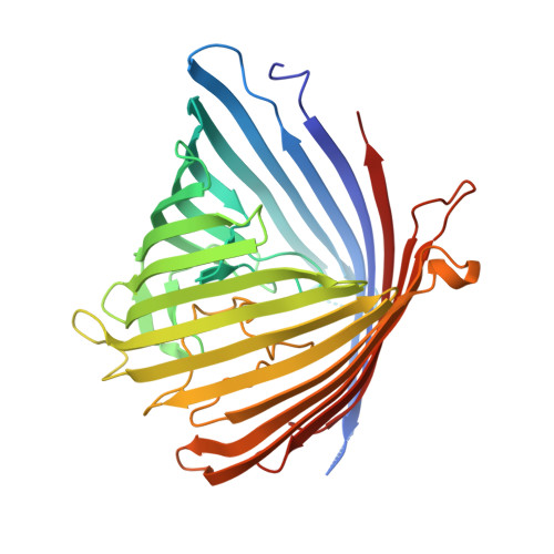
Toward Understanding the Outer Membrane Uptake of Small Molecules by Pseudomonas aeruginosa.

Eren, E.Parkin, J.Adelanwa, A.Cheneke, B.Movileanu, L.Khalid, S.van den Berg, B.

(2013) J Biol Chem 288: 12042-12053

  • DOI: https://doi.org/10.1074/jbc.M113.463570
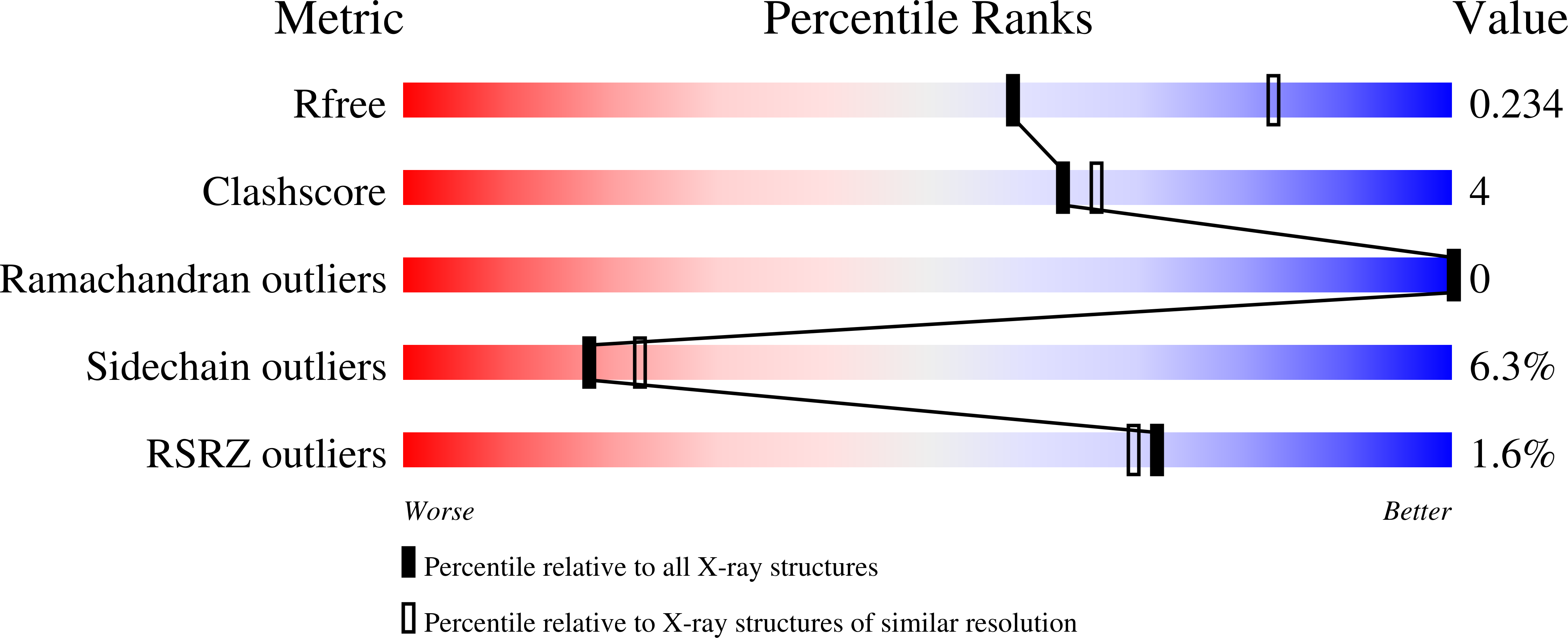
  • Primary Citation of Related Structures:  
    4FMS, 4FOZ

  • PubMed Abstract: 

    Because small molecules enter Gram-negative bacteria via outer membrane (OM) channels, understanding OM transport is essential for the rational design of improved and new antibiotics. In the human pathogen Pseudomonas aeruginosa, most small molecules are taken up by outer membrane carboxylate channel (Occ) proteins, which can be divided into two distinct subfamilies, OccD and OccK. Here we characterize substrate transport mediated by Occ proteins belonging to both subfamilies. Based on the determination of the OccK2-glucuronate co-crystal structure, we identify the channel residues that are essential for substrate transport. We further show that the pore regions of the channels are rigid in the OccK subfamily and highly dynamic in the OccD subfamily. We also demonstrate that the substrate carboxylate group interacts with central residues of the basic ladder, a row of arginine and lysine residues that leads to and away from the binding site at the channel constriction. Moreover, the importance of the basic ladder residues corresponds to their degree of conservation. Finally, we apply the generated insights by converting the archetype of the entire family, OccD1, from a basic amino acid-specific channel into a channel with a preference for negatively charged amino acids.


  • Organizational Affiliation

    Program in Molecular Medicine, University of Massachusetts Medical School, Worcester, Massachusetts 01605, USA.


Macromolecules
Find similar proteins by:  (by identity cutoff)  |  3D Structure
Entity ID: 1
MoleculeChains Sequence LengthOrganismDetailsImage
Probable porinA [auth B],
B [auth A]
397Pseudomonas aeruginosa PAO1Mutation(s): 0 
Gene Names: PA0240
Membrane Entity: Yes 
UniProt
Find proteins for Q9I6P8 (Pseudomonas aeruginosa (strain ATCC 15692 / DSM 22644 / CIP 104116 / JCM 14847 / LMG 12228 / 1C / PRS 101 / PAO1))
Explore Q9I6P8 
Go to UniProtKB:  Q9I6P8
Entity Groups  
Sequence Clusters30% Identity50% Identity70% Identity90% Identity95% Identity100% Identity
UniProt GroupQ9I6P8
Sequence Annotations
Expand
  • Reference Sequence
Small Molecules
Ligands 4 Unique
IDChains Name / Formula / InChI Key2D Diagram3D Interactions
PGV
Query on PGV

Download Ideal Coordinates CCD File 
J [auth B],
Q [auth A],
R [auth A],
S [auth A]
(1R)-2-{[{[(2S)-2,3-DIHYDROXYPROPYL]OXY}(HYDROXY)PHOSPHORYL]OXY}-1-[(PALMITOYLOXY)METHYL]ETHYL (11E)-OCTADEC-11-ENOATE
C40 H77 O10 P
ADYWCMPUNIVOEA-GPJPVTGXSA-N
C8E
Query on C8E

Download Ideal Coordinates CCD File 
F [auth B]
G [auth B]
H [auth B]
I [auth B]
N [auth A]
F [auth B],
G [auth B],
H [auth B],
I [auth B],
N [auth A],
O [auth A],
P [auth A]
(HYDROXYETHYLOXY)TRI(ETHYLOXY)OCTANE
C16 H34 O5
FEOZZFHAVXYAMB-UHFFFAOYSA-N
BDP
Query on BDP

Download Ideal Coordinates CCD File 
C [auth B],
K [auth A]
beta-D-glucopyranuronic acid
C6 H10 O7
AEMOLEFTQBMNLQ-QIUUJYRFSA-N
SO4
Query on SO4

Download Ideal Coordinates CCD File 
D [auth B],
E [auth B],
L [auth A],
M [auth A]
SULFATE ION
O4 S
QAOWNCQODCNURD-UHFFFAOYSA-L
Experimental Data & Validation

Experimental Data

  • Method: X-RAY DIFFRACTION
  • Resolution: 2.45 Å
  • R-Value Free: 0.242 
  • R-Value Work: 0.191 
  • R-Value Observed: 0.194 
  • Space Group: P 1 21 1
Unit Cell:
Length ( Å )Angle ( ˚ )
a = 46.435α = 90
b = 206.802β = 99.09
c = 51.747γ = 90
Software Package:
Software NamePurpose
HKL-2000data collection
PHASERphasing
PHENIXrefinement
HKL-2000data reduction
HKL-2000data scaling

Structure Validation

View Full Validation Report


Ligand Structure Quality Assessment 


Entry History 

Deposition Data

Revision History  (Full details and data files)

  • Version 1.0: 2013-03-13
    Type: Initial release
  • Version 1.1: 2013-03-27
    Changes: Database references
  • Version 1.2: 2013-05-22
    Changes: Database references
  • Version 1.3: 2020-07-29
    Type: Remediation
    Reason: Carbohydrate remediation
    Changes: Data collection, Derived calculations, Structure summary
  • Version 1.4: 2024-02-28
    Changes: Data collection, Database references, Structure summary