4FHT

Crystal Structure of the PcaV transcriptional regulator from Streptomyces coelicolor in complex with its natural ligand


Experimental Data Snapshot

  • Method: X-RAY DIFFRACTION
  • Resolution: 2.15 Å
  • R-Value Free: 0.244 
  • R-Value Work: 0.197 
  • R-Value Observed: 0.202 

wwPDB Validation   3D Report Full Report


Ligand Structure Quality Assessment 


This is version 1.1 of the entry. See complete history


Literature

Study of PcaV from Streptomyces coelicolor yields new insights into ligand-responsive MarR family transcription factors.

Davis, J.R.Brown, B.L.Page, R.Sello, J.K.

(2013) Nucleic Acids Res 41: 3888-3900

  • DOI: https://doi.org/10.1093/nar/gkt009
  • Primary Citation of Related Structures:  
    4FHT, 4G9Y

  • PubMed Abstract: 

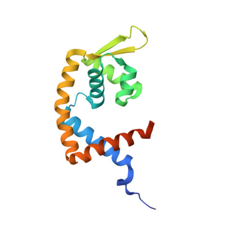
    MarR family proteins constitute a group of >12 000 transcriptional regulators encoded in bacterial and archaeal genomes that control gene expression in metabolism, stress responses, virulence and multi-drug resistance. There is much interest in defining the molecular mechanism by which ligand binding attenuates the DNA-binding activities of these proteins. Here, we describe how PcaV, a MarR family regulator in Streptomyces coelicolor, controls transcription of genes encoding β-ketoadipate pathway enzymes through its interaction with the pathway substrate, protocatechuate. This transcriptional repressor is the only MarR protein known to regulate this essential pathway for aromatic catabolism. In in vitro assays, protocatechuate and other phenolic compounds disrupt the PcaV-DNA complex. We show that PcaV binds protocatechuate in a 1:1 stoichiometry with the highest affinity of any MarR family member. Moreover, we report structures of PcaV in its apo form and in complex with protocatechuate. We identify an arginine residue that is critical for ligand coordination and demonstrate that it is also required for binding DNA. We propose that interaction of ligand with this arginine residue dictates conformational changes that modulate DNA binding. Our results provide new insights into the molecular mechanism by which ligands attenuate DNA binding in this large family of transcription factors.


  • Organizational Affiliation

    Department of Molecular Pharmacology and Physiology, Brown University, Providence, RI 02912, USA.


Macromolecules
Find similar proteins by:  (by identity cutoff)  |  3D Structure
Entity ID: 1
MoleculeChains Sequence LengthOrganismDetailsImage
PcaV transcriptional regulator
A, B
157Streptomyces coelicolor A3(2)Mutation(s): 0 
Gene Names: SCO6704
UniProt
Find proteins for Q9XAM6 (Streptomyces coelicolor (strain ATCC BAA-471 / A3(2) / M145))
Explore Q9XAM6 
Go to UniProtKB:  Q9XAM6
Entity Groups  
Sequence Clusters30% Identity50% Identity70% Identity90% Identity95% Identity100% Identity
UniProt GroupQ9XAM6
Sequence Annotations
Expand
  • Reference Sequence
Small Molecules
Binding Affinity Annotations 
IDSourceBinding Affinity
DHB Binding MOAD:  4FHT Kd: 670 (nM) from 1 assay(s)
PDBBind:  4FHT Kd: 670 (nM) from 1 assay(s)
Experimental Data & Validation

Experimental Data

  • Method: X-RAY DIFFRACTION
  • Resolution: 2.15 Å
  • R-Value Free: 0.244 
  • R-Value Work: 0.197 
  • R-Value Observed: 0.202 
  • Space Group: P 21 21 21
Unit Cell:
Length ( Å )Angle ( ˚ )
a = 62.357α = 90
b = 63.965β = 90
c = 73.009γ = 90
Software Package:
Software NamePurpose
CBASSdata collection
PHASERphasing
PHENIXrefinement
DENZOdata reduction
SCALEPACKdata scaling

Structure Validation

View Full Validation Report


Ligand Structure Quality Assessment 


Entry History 

Deposition Data

Revision History  (Full details and data files)

  • Version 1.0: 2013-04-24
    Type: Initial release
  • Version 1.1: 2023-09-13
    Changes: Data collection, Database references, Derived calculations, Refinement description