4F96

Crystal Structure of VldE, the pseudo-glycosyltransferase, in complex with GDP


Experimental Data Snapshot

  • Method: X-RAY DIFFRACTION
  • Resolution: 2.15 Å
  • R-Value Free: 0.209 
  • R-Value Work: 0.190 
  • R-Value Observed: 0.191 

wwPDB Validation   3D Report Full Report


Ligand Structure Quality Assessment 


This is version 1.1 of the entry. See complete history


Literature

Mechanistic Insights into Validoxylamine A 7'-Phosphate Synthesis by VldE Using the Structure of the Entire Product Complex.

Cavalier, M.C.Yim, Y.S.Asamizu, S.Neau, D.Almabruk, K.H.Mahmud, T.Lee, Y.H.

(2012) PLoS One 7: e44934-e44934

  • DOI: https://doi.org/10.1371/journal.pone.0044934
  • Primary Citation of Related Structures:  
    4F96, 4F97, 4F9F

  • PubMed Abstract: 

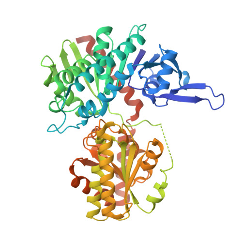
    The pseudo-glycosyltransferase VldE catalyzes non-glycosidic C-N coupling between an unsaturated cyclitol and a saturated aminocyclitol with the conservation of the stereochemical configuration of the substrates to form validoxylamine A 7'-phosphate, the biosynthetic precursor of the antibiotic validamycin A. To study the molecular basis of its mechanism, the three-dimensional structures of VldE from Streptomyces hygroscopicus subsp. limoneus was determined in apo form, in complex with GDP, in complex with GDP and validoxylamine A 7'-phosphate, and in complex with GDP and trehalose. The structure of VldE with the catalytic site in both an "open" and "closed" conformation is also described. With these structures, the preferred binding of the guanine moiety by VldE, rather than the uracil moiety as seen in OtsA could be explained. The elucidation of the VldE structure in complex with the entirety of its products provides insight into the internal return mechanism by which catalysis occurs with a net retention of the stereochemical configuration of the donated cyclitol.


  • Organizational Affiliation

    Department of Biological Sciences, Louisiana State University, Baton Rouge, Louisiana, United States of America.


Macromolecules
Find similar proteins by:  (by identity cutoff)  |  3D Structure
Entity ID: 1
MoleculeChains Sequence LengthOrganismDetailsImage
VldEA [auth B],
B [auth A]
497Streptomyces hygroscopicus subsp. limoneusMutation(s): 0 
Gene Names: vldE
EC: 2.4
UniProt
Find proteins for Q15JG1 (Streptomyces hygroscopicus subsp. limoneus)
Explore Q15JG1 
Go to UniProtKB:  Q15JG1
Entity Groups  
Sequence Clusters30% Identity50% Identity70% Identity90% Identity95% Identity100% Identity
UniProt GroupQ15JG1
Sequence Annotations
Expand
  • Reference Sequence
Experimental Data & Validation

Experimental Data

  • Method: X-RAY DIFFRACTION
  • Resolution: 2.15 Å
  • R-Value Free: 0.209 
  • R-Value Work: 0.190 
  • R-Value Observed: 0.191 
  • Space Group: P 1 21 1
Unit Cell:
Length ( Å )Angle ( ˚ )
a = 47.821α = 90
b = 121.151β = 91.74
c = 84.333γ = 90
Software Package:
Software NamePurpose
HKL-2000data collection
PHENIXmodel building
PHENIXrefinement
HKL-2000data reduction
HKL-2000data scaling
PHENIXphasing

Structure Validation

View Full Validation Report


Ligand Structure Quality Assessment 


Entry History 

Deposition Data

Revision History  (Full details and data files)

  • Version 1.0: 2012-10-17
    Type: Initial release
  • Version 1.1: 2024-02-28
    Changes: Data collection, Database references, Derived calculations