


Crystal Structure of the alpha spectrin SH3 domain at pH 5
Camara-Artigas, A., Gavira, J.A.To be published.
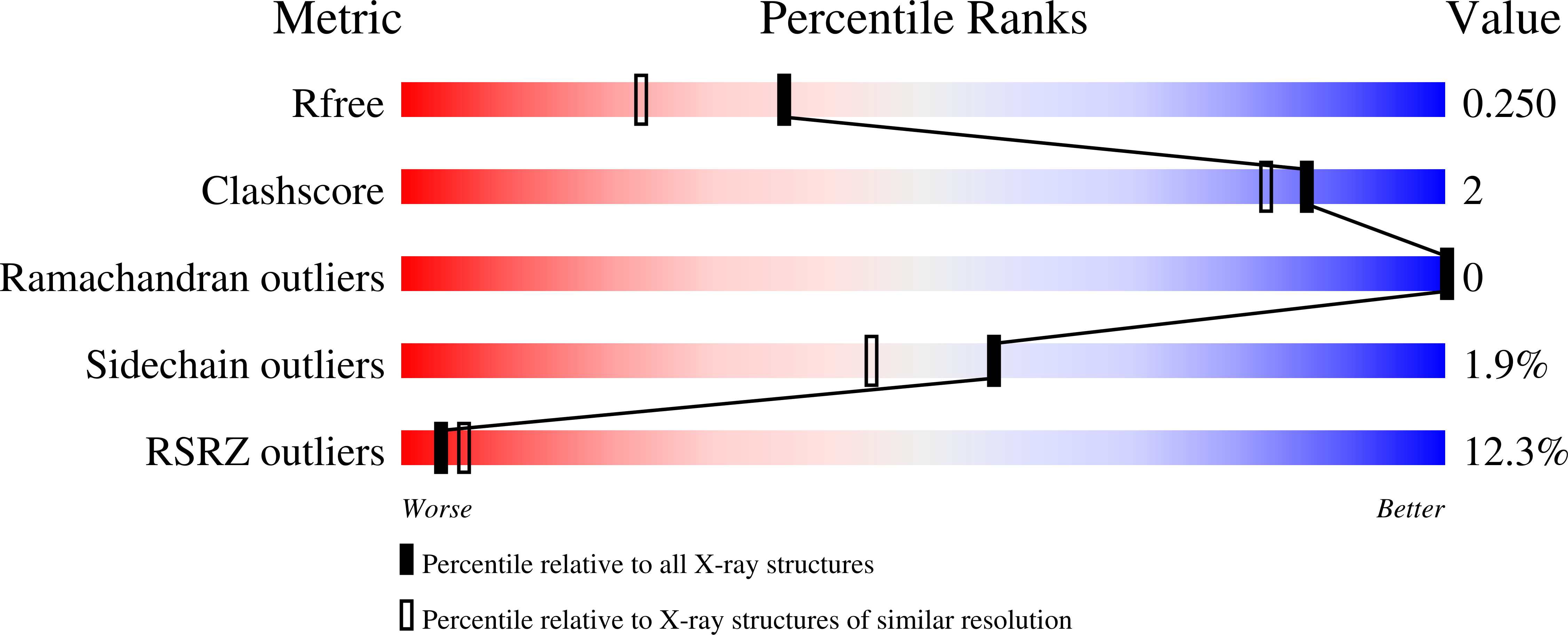
Experimental Data Snapshot
wwPDB Validation 3D Report Full Report
Entity ID: 1 | |||||
|---|---|---|---|---|---|
| Molecule | Chains | Sequence Length | Organism | Details | Image |
| Spectrin alpha chain, brain | 62 | Gallus gallus | Mutation(s): 0 Gene Names: SPTAN1, SPTA2 | 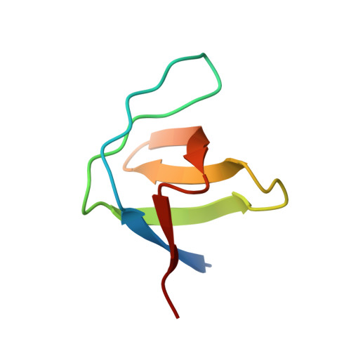 | |
UniProt | |||||
Find proteins for P07751 (Gallus gallus) Explore P07751 Go to UniProtKB: P07751 | |||||
Entity Groups | |||||
| Sequence Clusters | 30% Identity50% Identity70% Identity90% Identity95% Identity100% Identity | ||||
| UniProt Group | P07751 | ||||
Sequence AnnotationsExpand | |||||
| |||||
| Length ( Å ) | Angle ( ˚ ) |
|---|---|
| a = 32.72 | α = 90 |
| b = 42.336 | β = 90 |
| c = 50.335 | γ = 90 |
| Software Name | Purpose |
|---|---|
| DENZO | data reduction |
| SCALA | data scaling |
| MOLREP | phasing |
| PHENIX | refinement |
| PDB_EXTRACT | data extraction |
| SCALEPACK | data scaling |
RCSB PDB (citation) is hosted by
RCSB PDB is a member of the

