New Aspects of the Phosphatase VHZ Revealed by a High-Resolution Structure with Vanadate and Substrate Screening.
Kuznetsov, V.I., Hengge, A.C., Johnson, S.J.(2012) Biochemistry 51: 9869-9879
- PubMed: 23145819
- DOI: https://doi.org/10.1021/bi300908y
- Primary Citation of Related Structures:
4ERC - PubMed Abstract:
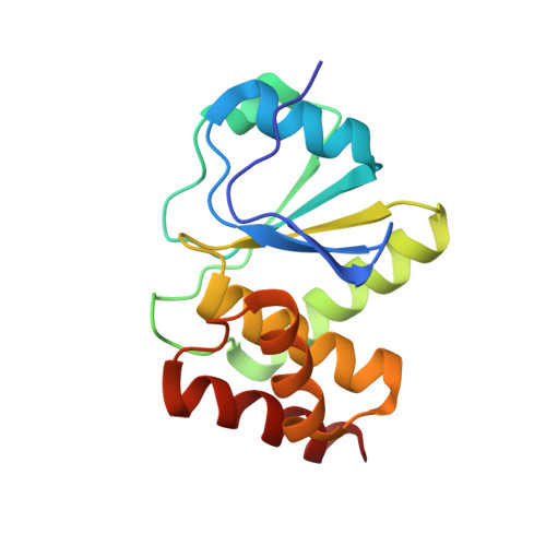
The recently discovered 150-residue human VHZ (VH1-related protein, Z member) is one of the smallest protein tyrosine phosphatases (PTPs) known and contains only the minimal structural elements common to all PTPs. We report a substrate screening analysis and a crystal structure of the VHZ complex with vanadate at 1.1 Å resolution, with a detailed structural comparison with other members of the protein tyrosine phosphatase family, including classical tyrosine-specific protein tyrosine phosphatases (PTPs) and dual-specificity phosphatases (DSPs). A screen with 360 phosphorylated peptides shows VHZ efficiently catalyzes the hydrolysis of phosphotyrosine (pY)-containing peptides but exhibits no activity toward phosphoserine (pS) or phosphothreonine (pT) peptides. The new structure reveals a deep and narrow active site more typical of the classical tyrosine-specific PTPs. Despite the high degrees of structural and sequence similarity between VHZ and classical PTPs, its general acid IPD-loop is most likely conformationally rigid, in contrast to the flexible WPD counterpart of classical PTPs. VHZ also lacks substrate recognition domains and other domains typically found on classical PTPs. It is therefore proposed that VHZ is more properly classified as an atypical PTP rather than an atypical DSP, as has been suggested.
Organizational Affiliation:
Department of Chemistry and Biochemistry, Utah State University, Logan, UT 84322-0300, USA.