4EPC

Crystal structure of Autolysin repeat domains from Staphylococcus epidermidis


Experimental Data Snapshot

  • Method: X-RAY DIFFRACTION
  • Resolution: 2.90 Å
  • R-Value Free: 0.298 
  • R-Value Work: 0.263 
  • R-Value Observed: 0.266 

wwPDB Validation   3D Report Full Report


This is version 1.1 of the entry. See complete history


Literature

Ligand-binding properties and conformational dynamics of autolysin repeat domains in staphylococcal cell wall recognition.

Zoll, S.Schlag, M.Shkumatov, A.V.Rautenberg, M.Svergun, D.I.Gotz, F.Stehle, T.

(2012) J Bacteriol 194: 3789-3802

  • DOI: https://doi.org/10.1128/JB.00331-12
  • Primary Citation of Related Structures:  
    4EPC

  • PubMed Abstract: 

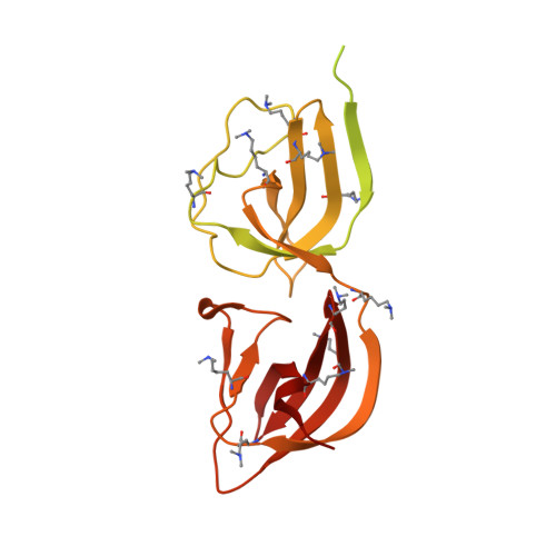
    The bifunctional major autolysin Atl plays a key role in staphylococcal cell separation. Processing of Atl yields catalytically active amidase (AM) and glucosaminidase (GL) domains that are each fused to repeating units. The two repeats of AM (R1 and R2) target the enzyme to the septum, where it cleaves murein between dividing cells. We have determined the crystal structure of R2, which reveals that each repeat folds into two half-open β-barrel subunits. We further demonstrate that lipoteichoic acid serves as a receptor for the repeats and that this interaction depends on conserved surfaces in each subunit. Small-angle X-ray scattering of the mature amidase reveals the presence of flexible linkers separating the AM, R1, and R2 units. Different levels of flexibility for each linker provide mechanistic insights into the conformational dynamics of the full-length protein and the roles of its components in cell wall association and catalysis. Our analysis supports a model in which the repeats direct the catalytic AM domain to the septum, where it can optimally perform the final step of cell division.


  • Organizational Affiliation

    Interfaculty Institute of Biochemistry, University of Tuebingen, Tuebingen, Germany.


Macromolecules
Find similar proteins by:  (by identity cutoff)  |  3D Structure
Entity ID: 1
MoleculeChains Sequence LengthOrganismDetailsImage
N-acetylmuramoyl-L-alanine amidase334Staphylococcus epidermidisMutation(s): 0 
Gene Names: atlatlE
EC: 3.5.1.28
UniProt
Find proteins for O33635 (Staphylococcus epidermidis)
Explore O33635 
Go to UniProtKB:  O33635
Entity Groups  
Sequence Clusters30% Identity50% Identity70% Identity90% Identity95% Identity100% Identity
UniProt GroupO33635
Sequence Annotations
Expand
  • Reference Sequence
Small Molecules
Modified Residues  1 Unique
IDChains TypeFormula2D DiagramParent
MLY
Query on MLY
A
L-PEPTIDE LINKINGC8 H18 N2 O2LYS
Experimental Data & Validation

Experimental Data

  • Method: X-RAY DIFFRACTION
  • Resolution: 2.90 Å
  • R-Value Free: 0.298 
  • R-Value Work: 0.263 
  • R-Value Observed: 0.266 
  • Space Group: P 61 2 2
Unit Cell:
Length ( Å )Angle ( ˚ )
a = 95.36α = 90
b = 95.36β = 90
c = 233.67γ = 120
Software Package:
Software NamePurpose
XDSdata scaling
SOLVEphasing
BUSTERrefinement
XDSdata reduction

Structure Validation

View Full Validation Report


Entry History 

Deposition Data

Revision History  (Full details and data files)

  • Version 1.0: 2012-06-06
    Type: Initial release
  • Version 1.1: 2012-08-15
    Changes: Database references