


Crystal Structure of the Putative acetyltransferase from Streptococcus mutans
Li, G.L., Nie, J.K., Li, L.F., Su, X.D.To be published.
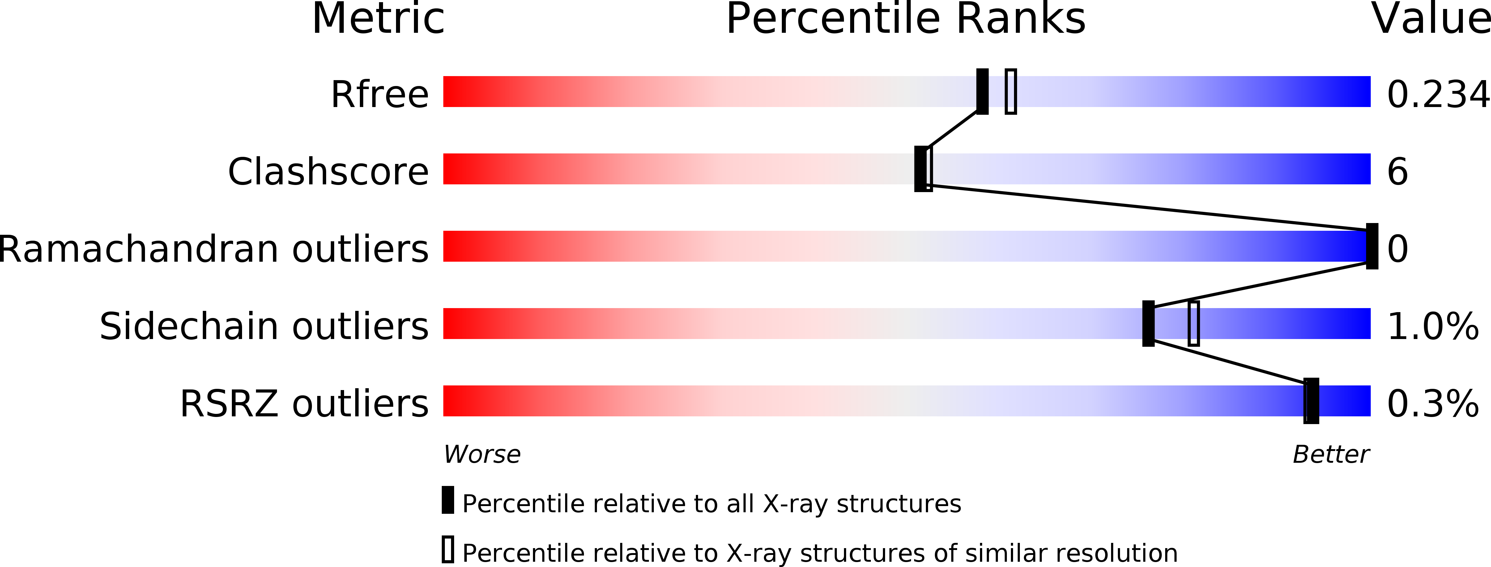
Experimental Data Snapshot
wwPDB Validation 3D Report Full Report
Entity ID: 1 | |||||
|---|---|---|---|---|---|
| Molecule | Chains | Sequence Length | Organism | Details | Image |
| Putative acetyltransferase | 207 | Streptococcus mutans | Mutation(s): 0 Gene Names: SMU_1511c | 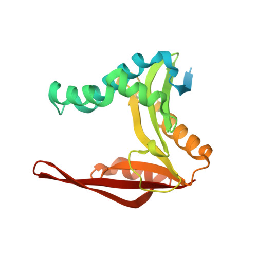 | |
UniProt | |||||
Find proteins for Q8DT67 (Streptococcus mutans serotype c (strain ATCC 700610 / UA159)) Explore Q8DT67 Go to UniProtKB: Q8DT67 | |||||
Entity Groups | |||||
| Sequence Clusters | 30% Identity50% Identity70% Identity90% Identity95% Identity100% Identity | ||||
| UniProt Group | Q8DT67 | ||||
Sequence AnnotationsExpand | |||||
| |||||
| Length ( Å ) | Angle ( ˚ ) |
|---|---|
| a = 75.7 | α = 90 |
| b = 75.7 | β = 90 |
| c = 80.9 | γ = 90 |
| Software Name | Purpose |
|---|---|
| ADSC | data collection |
| PHENIX | model building |
| PHENIX | refinement |
| XDS | data reduction |
| XSCALE | data scaling |
| PHENIX | phasing |
RCSB PDB (citation) is hosted by
RCSB PDB is a member of the

