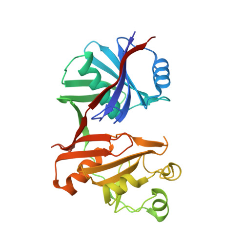
1.76A X-ray Crystal Structure of a Putative Phenazine Biosynthesis PhzC/PhzF Protein from Clostridium difficile (strain 630)
Brunzelle, J.S., Wawrzak, W., Kudritska, M., Anderson, W.F., Savchenko, A., Center for Structural Genomics of Infectious Diseases (CSGID)To be published.