4DDL

PDE10a Crystal Structure Complexed with Novel Inhibitor


Experimental Data Snapshot

  • Method: X-RAY DIFFRACTION
  • Resolution: 2.07 Å
  • R-Value Free: 0.195 
  • R-Value Work: 0.186 
  • R-Value Observed: 0.186 

wwPDB Validation   3D Report Full Report


Ligand Structure Quality Assessment 


This is version 1.0 of the entry. See complete history


Literature

Discovery of potent, selective, and metabolically stable 4-(pyridin-3-yl)cinnolines as novel phosphodiesterase 10A (PDE10A) inhibitors.

Hu, E.Kunz, R.K.Rumfelt, S.Chen, N.Burli, R.Li, C.Andrews, K.L.Zhang, J.Chmait, S.Kogan, J.Lindstrom, M.Hitchcock, S.A.Treanor, J.

(2012) Bioorg Med Chem Lett 22: 2262-2265

  • DOI: https://doi.org/10.1016/j.bmcl.2012.01.086
  • Primary Citation of Related Structures:  
    4DDL

  • PubMed Abstract: 

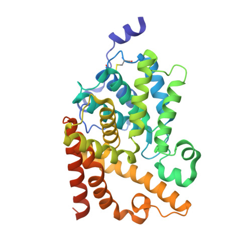
    We report the discovery of 6,7-dimethoxy-4-(pyridin-3-yl)cinnolines as novel inhibitors of phosphodiesterase 10A (PDE10A). Systematic examination and analyses of structure-activity-relationships resulted in single digit nM potency against PDE10A. X-ray co-crystal structure revealed the mode of binding in the enzyme's catalytic domain and the source of selectivity against other PDEs. High in vivo clearance in rats was addressed with the help of metabolite identification (ID) studies. These findings combined resulted in compound 39, a promising potent inhibitor of PDE10A with good in vivo metabolic stability in rats and efficacy in a rodent behavioral model.


  • Organizational Affiliation

    Department of Small Molecule Chemistry, Amgen Inc., Thousand Oaks, CA 91320-1799, USA.


Macromolecules
Find similar proteins by:  (by identity cutoff)  |  3D Structure
Entity ID: 1
MoleculeChains Sequence LengthOrganismDetailsImage
cAMP and cAMP-inhibited cGMP 3',5'-cyclic phosphodiesterase 10A
A, B
338Homo sapiensMutation(s): 0 
Gene Names: PDE10A
EC: 3.1.4.17 (PDB Primary Data), 3.1.4.35 (PDB Primary Data)
UniProt & NIH Common Fund Data Resources
Find proteins for Q9Y233 (Homo sapiens)
Explore Q9Y233 
Go to UniProtKB:  Q9Y233
PHAROS:  Q9Y233
GTEx:  ENSG00000112541 
Entity Groups  
Sequence Clusters30% Identity50% Identity70% Identity90% Identity95% Identity100% Identity
UniProt GroupQ9Y233
Sequence Annotations
Expand
  • Reference Sequence
Small Molecules
Ligands 3 Unique
IDChains Name / Formula / InChI Key2D Diagram3D Interactions
0JQ
Query on 0JQ

Download Ideal Coordinates CCD File 
L [auth A],
W [auth B]
2-{1-[5-(6,7-dimethoxycinnolin-4-yl)-3-methylpyridin-2-yl]piperidin-4-yl}propan-2-ol
C24 H30 N4 O3
PPAWFHDEPAUENY-UHFFFAOYSA-N
SO4
Query on SO4

Download Ideal Coordinates CCD File 
E [auth A]
F [auth A]
G [auth A]
H [auth A]
I [auth A]
E [auth A],
F [auth A],
G [auth A],
H [auth A],
I [auth A],
J [auth A],
K [auth A],
O [auth B],
P [auth B],
Q [auth B],
R [auth B],
S [auth B],
T [auth B],
U [auth B],
V [auth B]
SULFATE ION
O4 S
QAOWNCQODCNURD-UHFFFAOYSA-L
ZN
Query on ZN

Download Ideal Coordinates CCD File 
C [auth A],
D [auth A],
M [auth B],
N [auth B]
ZINC ION
Zn
PTFCDOFLOPIGGS-UHFFFAOYSA-N
Binding Affinity Annotations 
IDSourceBinding Affinity
0JQ BindingDB:  4DDL IC50: 4.9 (nM) from 1 assay(s)
PDBBind:  4DDL IC50: 4.9 (nM) from 1 assay(s)
Binding MOAD:  4DDL IC50: 4.9 (nM) from 1 assay(s)
Experimental Data & Validation

Experimental Data

  • Method: X-RAY DIFFRACTION
  • Resolution: 2.07 Å
  • R-Value Free: 0.195 
  • R-Value Work: 0.186 
  • R-Value Observed: 0.186 
  • Space Group: F 2 3
Unit Cell:
Length ( Å )Angle ( ˚ )
a = 252.964α = 90
b = 252.964β = 90
c = 252.964γ = 90
Software Package:
Software NamePurpose
StructureStudiodata collection
REFMACrefinement
HKL-2000data reduction
HKL-2000data scaling

Structure Validation

View Full Validation Report


Ligand Structure Quality Assessment 


Entry History 

Deposition Data

Revision History  (Full details and data files)

  • Version 1.0: 2012-03-21
    Type: Initial release