4D60

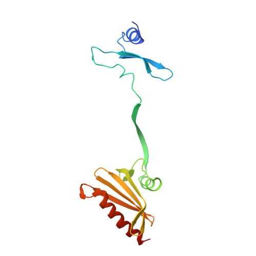
Structure of a dimeric Plasmodium falciparum profilin mutant


Experimental Data Snapshot

  • Method: X-RAY DIFFRACTION
  • Resolution: 3.30 Å
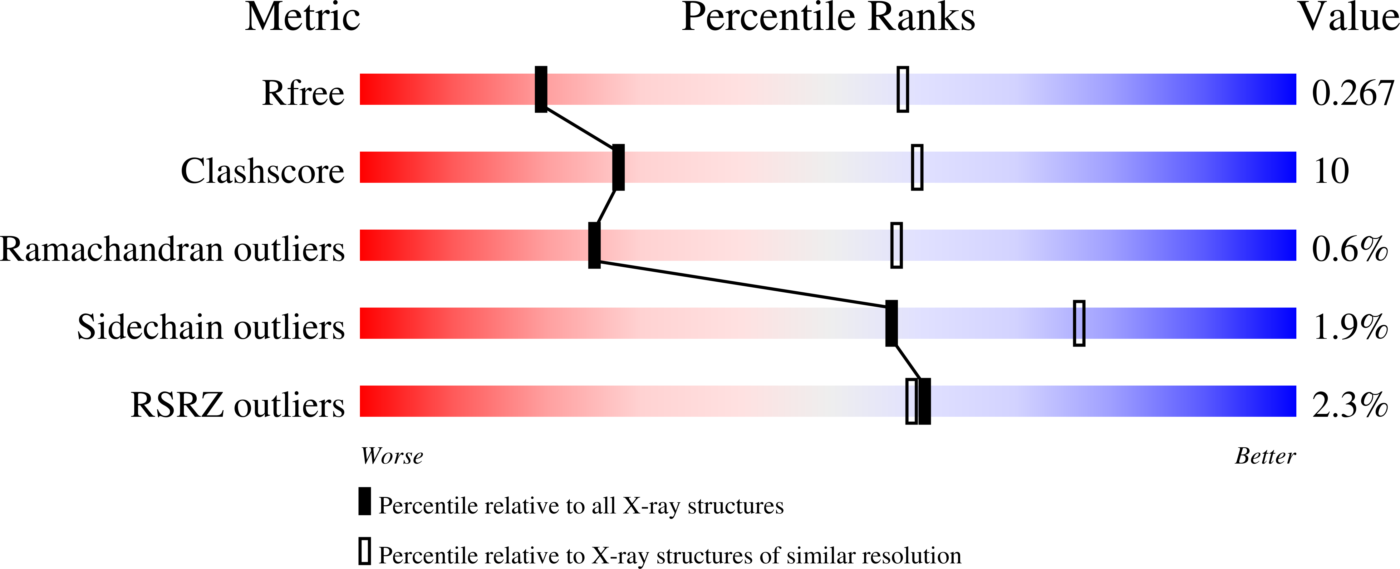
  • R-Value Free: 0.263 
  • R-Value Work: 0.240 
  • R-Value Observed: 0.241 

wwPDB Validation   3D Report Full Report


This is version 1.4 of the entry. See complete history


Literature

Two Independently Folding Units of Plasmodium Profilin Suggest Evolution Via Gene Fusion.

Bhargav, S.P.Vahokoski, J.Kallio, J.P.Torda, A.E.Kursula, P.Kursula, I.

(2015) Cell Mol Life Sci 72: 4193

  • DOI: https://doi.org/10.1007/s00018-015-1932-0
  • Primary Citation of Related Structures:  
    4D60

  • PubMed Abstract: 

    Gene fusion is a common mechanism of protein evolution that has mainly been discussed in the context of multidomain or symmetric proteins. Less is known about fusion of ancestral genes to produce small single-domain proteins. Here, we show with a domain-swapped mutant Plasmodium profilin that this small, globular, apparently single-domain protein consists of two foldons. The separation of binding sites for different protein ligands in the two halves suggests evolution via an ancient gene fusion event, analogous to the formation of multidomain proteins. Finally, the two fragments can be assembled together after expression as two separate gene products. The possibility to engineer both domain-swapped dimers and half-profilins that can be assembled back to a full profilin provides perspectives for engineering of novel protein folds, e.g., with different scaffolding functions.


  • Organizational Affiliation

    Faculty of Biochemistry and Molecular Medicine, University of Oulu, P.O. Box 5400, 90014, Oulu, Finland.


Macromolecules
Find similar proteins by:  (by identity cutoff)  |  3D Structure
Entity ID: 1
MoleculeChains Sequence LengthOrganismDetailsImage
PROFILIN
A, B, C, D, E
A, B, C, D, E, F, G, H, I, J, K, L, M, N, O, P
169Plasmodium falciparumMutation(s): 0 
UniProt
Find proteins for Q8I2J4 (Plasmodium falciparum (isolate 3D7))
Explore Q8I2J4 
Go to UniProtKB:  Q8I2J4
Entity Groups  
Sequence Clusters30% Identity50% Identity70% Identity90% Identity95% Identity100% Identity
UniProt GroupQ8I2J4
Sequence Annotations
Expand
  • Reference Sequence
Experimental Data & Validation

Experimental Data

  • Method: X-RAY DIFFRACTION
  • Resolution: 3.30 Å
  • R-Value Free: 0.263 
  • R-Value Work: 0.240 
  • R-Value Observed: 0.241 
  • Space Group: P 21 21 21
Unit Cell:
Length ( Å )Angle ( ˚ )
a = 84.07α = 90
b = 246.46β = 90
c = 256.74γ = 90
Software Package:
Software NamePurpose
PHENIXrefinement
XDSdata reduction
XSCALEdata scaling
PHASERphasing

Structure Validation

View Full Validation Report


Entry History 

Deposition Data

Revision History  (Full details and data files)

  • Version 1.0: 2015-06-03
    Type: Initial release
  • Version 1.1: 2015-06-10
    Changes: Data collection, Database references
  • Version 1.2: 2015-10-28
    Changes: Database references
  • Version 1.3: 2018-03-07
    Changes: Data collection, Source and taxonomy
  • Version 1.4: 2023-12-20
    Changes: Data collection, Database references, Derived calculations, Other, Refinement description