4CVB

Crystal structure of quinone-dependent alcohol dehydrogenase from Pseudogluconobacter saccharoketogenenes


Experimental Data Snapshot

  • Method: X-RAY DIFFRACTION
  • Resolution: 1.72 Å
  • R-Value Free: 0.149 
  • R-Value Work: 0.128 
  • R-Value Observed: 0.129 

wwPDB Validation   3D Report Full Report


Ligand Structure Quality Assessment 


This is version 1.4 of the entry. See complete history


Literature

Crystal Structure of Quinone-Dependent Alcohol Dehydrogenase from Pseudogluconobacter Saccharoketogenes. A Versatile Dehydrogenase Oxidizing Alcohols and Carbohydrates.

Rozeboom, H.J.Yu, S.Mikkelsen, R.Nikolaev, I.Mulder, H.J.Dijkstra, B.W.

(2015) Protein Sci 24: 2044

  • DOI: https://doi.org/10.1002/pro.2818
  • Primary Citation of Related Structures:  
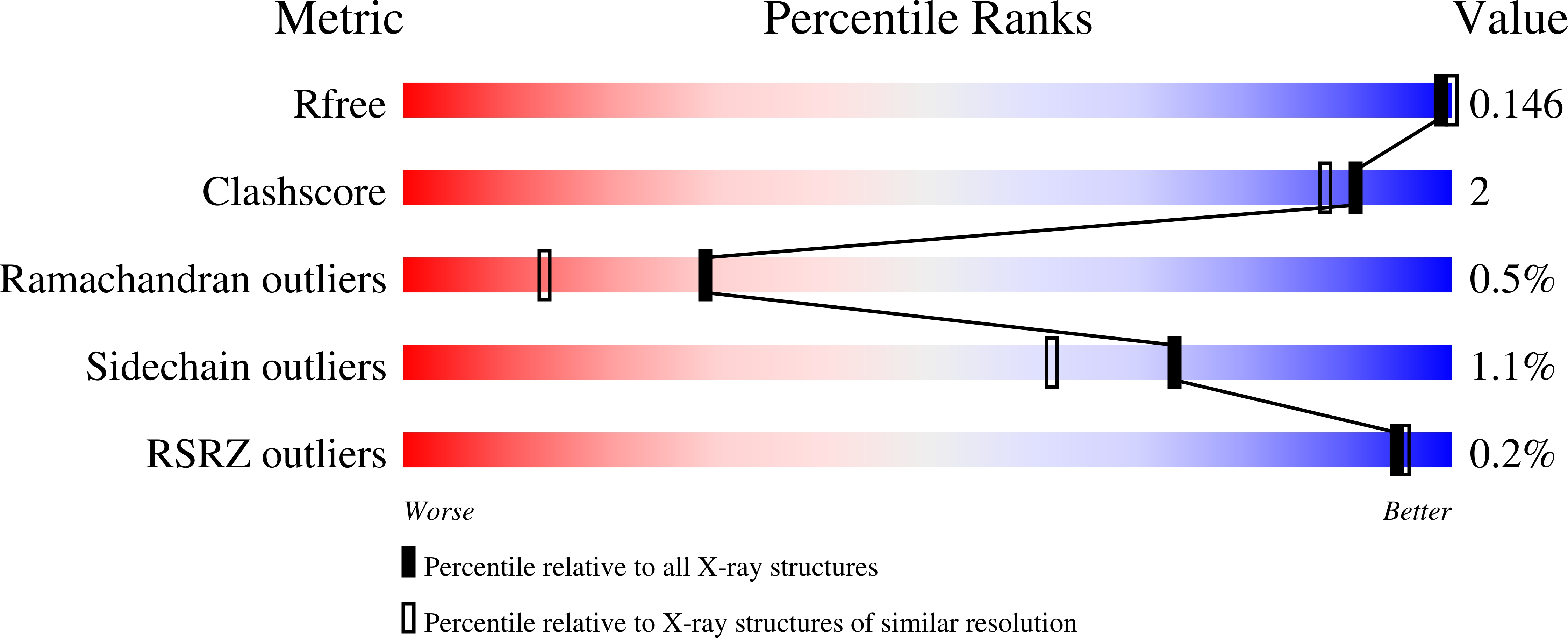
    4CVB, 4CVC

  • PubMed Abstract: 

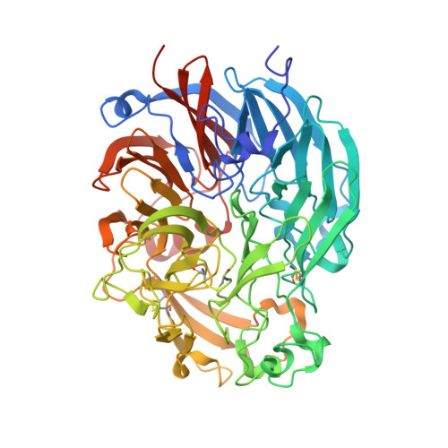
    The quinone-dependent alcohol dehydrogenase (PQQ-ADH, E.C. 1.1.5.2) from the Gram-negative bacterium Pseudogluconobacter saccharoketogenes IFO 14464 oxidizes primary alcohols (e.g. ethanol, butanol), secondary alcohols (monosaccharides), as well as aldehydes, polysaccharides, and cyclodextrins. The recombinant protein, expressed in Pichia pastoris, was crystallized, and three-dimensional (3D) structures of the native form, with PQQ and a Ca(2+) ion, and of the enzyme in complex with a Zn(2+) ion and a bound substrate mimic were determined at 1.72 Å and 1.84 Å resolution, respectively. PQQ-ADH displays an eight-bladed β-propeller fold, characteristic of Type I quinone-dependent methanol dehydrogenases. However, three of the four ligands of the Ca(2+) ion differ from those of related dehydrogenases and they come from different parts of the polypeptide chain. These differences result in a more open, easily accessible active site, which explains why PQQ-ADH can oxidize a broad range of substrates. The bound substrate mimic suggests Asp333 as the catalytic base. Remarkably, no vicinal disulfide bridge is present near the PQQ, which in other PQQ-dependent alcohol dehydrogenases has been proposed to be necessary for electron transfer. Instead an associated cytochrome c can approach the PQQ for direct electron transfer.


  • Organizational Affiliation

    Laboratory of Biophysical Chemistry, Groningen Biomolecular Sciences and Biotechnology Institute, University of Groningen, Groningen, The Netherlands.


Macromolecules
Find similar proteins by:  (by identity cutoff)  |  3D Structure
Entity ID: 1
MoleculeChains Sequence LengthOrganismDetailsImage
ALCOHOL DEHYDROGENASE572Pseudogluconobacter saccharoketogenesMutation(s): 0 
EC: 1.1.2.8
UniProt
Find proteins for Q93RE9 (Pseudogluconobacter saccharoketogenes)
Explore Q93RE9 
Go to UniProtKB:  Q93RE9
Entity Groups  
Sequence Clusters30% Identity50% Identity70% Identity90% Identity95% Identity100% Identity
UniProt GroupQ93RE9
Sequence Annotations
Expand
  • Reference Sequence
Small Molecules
Ligands 5 Unique
IDChains Name / Formula / InChI Key2D Diagram3D Interactions
PQQ
Query on PQQ

Download Ideal Coordinates CCD File 
C [auth A]PYRROLOQUINOLINE QUINONE
C14 H6 N2 O8
MMXZSJMASHPLLR-UHFFFAOYSA-N
PPI
Query on PPI

Download Ideal Coordinates CCD File 
I [auth A]PROPANOIC ACID
C3 H6 O2
XBDQKXXYIPTUBI-UHFFFAOYSA-N
ZN
Query on ZN

Download Ideal Coordinates CCD File 
D [auth A],
E [auth A],
F [auth A],
G [auth A]
ZINC ION
Zn
PTFCDOFLOPIGGS-UHFFFAOYSA-N
CA
Query on CA

Download Ideal Coordinates CCD File 
B [auth A]CALCIUM ION
Ca
BHPQYMZQTOCNFJ-UHFFFAOYSA-N
CL
Query on CL

Download Ideal Coordinates CCD File 
H [auth A]CHLORIDE ION
Cl
VEXZGXHMUGYJMC-UHFFFAOYSA-M
Experimental Data & Validation

Experimental Data

  • Method: X-RAY DIFFRACTION
  • Resolution: 1.72 Å
  • R-Value Free: 0.149 
  • R-Value Work: 0.128 
  • R-Value Observed: 0.129 
  • Space Group: C 1 2 1
Unit Cell:
Length ( Å )Angle ( ˚ )
a = 127.95α = 90
b = 87.19β = 90.16
c = 57.1γ = 90
Software Package:
Software NamePurpose
REFMACrefinement
iMOSFLMdata reduction
SCALAdata scaling
PHASERphasing

Structure Validation

View Full Validation Report


Ligand Structure Quality Assessment 


Entry History 

Deposition Data

Revision History  (Full details and data files)

  • Version 1.0: 2015-03-25
    Type: Initial release
  • Version 1.1: 2015-10-21
    Changes: Database references
  • Version 1.2: 2016-01-27
    Changes: Database references
  • Version 1.3: 2017-07-05
    Changes: Data collection
  • Version 1.4: 2023-12-20
    Changes: Data collection, Database references, Derived calculations, Other, Refinement description