4BWW

Crystal structure of spin labelled azurin T21R1.


Experimental Data Snapshot

  • Method: X-RAY DIFFRACTION
  • Resolution: 1.48 Å
  • R-Value Free: 0.210 
  • R-Value Work: 0.179 
  • R-Value Observed: 0.180 

wwPDB Validation   3D Report Full Report


This is version 1.1 of the entry. See complete history


Literature

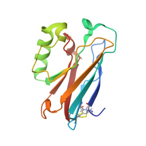
High-Resolution Crystal Structure of Spin Labelled (T21R1) Azurin from Pseudomonas Aeruginosa: A Challenging Structural Benchmark for in Silico Spin Labelling Algorithms.

Florin, N.Schiemann, O.Hagelueken, G.

(2014) BMC Struct Biol 14: 16

  • DOI: https://doi.org/10.1186/1472-6807-14-16
  • Primary Citation of Related Structures:  
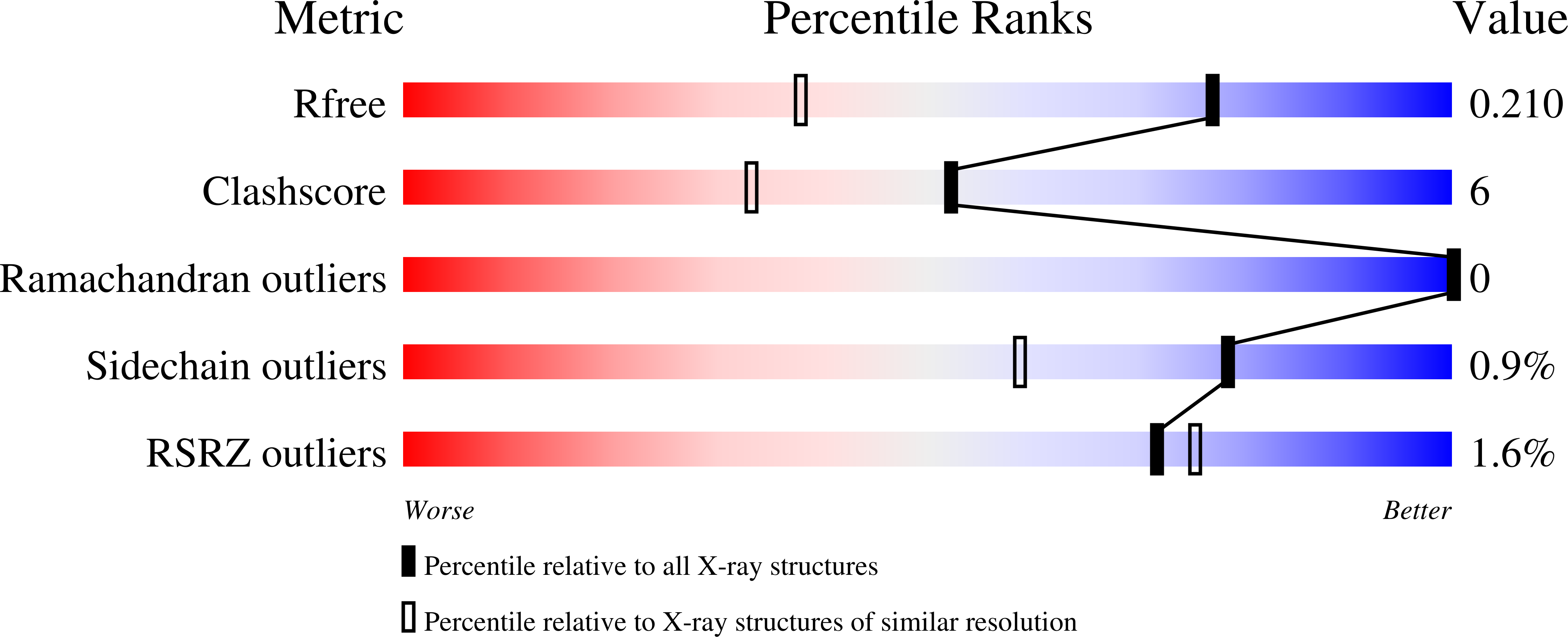
    4BWW

  • PubMed Abstract: 

    EPR-based distance measurements between spin labels in proteins have become a valuable tool in structural biology. The direct translation of the experimental distances into structural information is however often impaired by the intrinsic flexibility of the spin labelled side chains. Different algorithms exist that predict the approximate conformation of the spin label either by using pre-computed rotamer libraries of the labelled side chain (rotamer approach) or by simply determining its accessible volume (accessible volume approach). Surprisingly, comparisons with many experimental distances have shown that both approaches deliver the same distance prediction accuracy of about 3 Å.


  • Organizational Affiliation

    Institute for Physical and Theoretical Chemistry, University of Bonn, Wegelerstr, 12, Bonn, NRW 53115, Germany. hagelueken@pc.uni-bonn.de.


Macromolecules
Find similar proteins by:  (by identity cutoff)  |  3D Structure
Entity ID: 1
MoleculeChains Sequence LengthOrganismDetailsImage
AZURIN
A, B, C, D
131Pseudomonas aeruginosa PAO1Mutation(s): 1 
UniProt
Find proteins for P00282 (Pseudomonas aeruginosa (strain ATCC 15692 / DSM 22644 / CIP 104116 / JCM 14847 / LMG 12228 / 1C / PRS 101 / PAO1))
Explore P00282 
Go to UniProtKB:  P00282
Entity Groups  
Sequence Clusters30% Identity50% Identity70% Identity90% Identity95% Identity100% Identity
UniProt GroupP00282
Sequence Annotations
Expand
  • Reference Sequence
Small Molecules
Ligands 4 Unique
IDChains Name / Formula / InChI Key2D Diagram3D Interactions
SO4
Query on SO4

Download Ideal Coordinates CCD File 
L [auth B],
M [auth B],
T [auth C],
Y [auth D],
Z [auth D]
SULFATE ION
O4 S
QAOWNCQODCNURD-UHFFFAOYSA-L
GOL
Query on GOL

Download Ideal Coordinates CCD File 
F [auth A]
J [auth B]
K [auth B]
N [auth B]
P [auth B]
F [auth A],
J [auth B],
K [auth B],
N [auth B],
P [auth B],
R [auth C],
S [auth C],
W [auth D]
GLYCEROL
C3 H8 O3
PEDCQBHIVMGVHV-UHFFFAOYSA-N
CU
Query on CU

Download Ideal Coordinates CCD File 
E [auth A],
H [auth B],
Q [auth C],
V [auth D]
COPPER (II) ION
Cu
JPVYNHNXODAKFH-UHFFFAOYSA-N
NO3
Query on NO3

Download Ideal Coordinates CCD File 
G [auth A],
I [auth B],
O [auth B],
U [auth C],
X [auth D]
NITRATE ION
N O3
NHNBFGGVMKEFGY-UHFFFAOYSA-N
Modified Residues  1 Unique
IDChains TypeFormula2D DiagramParent
R1A
Query on R1A
A, B, C, D
L-PEPTIDE LINKINGC12 H21 N2 O3 S2CYS
Experimental Data & Validation

Experimental Data

  • Method: X-RAY DIFFRACTION
  • Resolution: 1.48 Å
  • R-Value Free: 0.210 
  • R-Value Work: 0.179 
  • R-Value Observed: 0.180 
  • Space Group: P 1
Unit Cell:
Length ( Å )Angle ( ˚ )
a = 36.95α = 74.4
b = 53.66β = 89.33
c = 73.21γ = 83.41
Software Package:
Software NamePurpose
PHENIXrefinement
iMOSFLMdata reduction
SCALAdata scaling
PHASERphasing

Structure Validation

View Full Validation Report


Entry History 

Deposition Data

Revision History  (Full details and data files)

  • Version 1.0: 2014-06-11
    Type: Initial release
  • Version 1.1: 2023-12-20
    Changes: Data collection, Database references, Derived calculations, Other, Refinement description