


The Structure of B. Circulans Aman6 Defines the Fold and Mechanism of the Gh-76 Mannoprotein Transglycosylases and Bacterial Mannosidases
Striebeck, A., Borodkin, V.S., Ferenbach, A.T., Van Aalten, D.M.F.To be published.
Experimental Data Snapshot
wwPDB Validation 3D Report Full Report

Entity ID: 1 | |||||
|---|---|---|---|---|---|
| Molecule | Chains | Sequence Length | Organism | Details | Image |
| ALPHA-1,6-MANNANASE | 341 | Niallia circulans | Mutation(s): 0 | 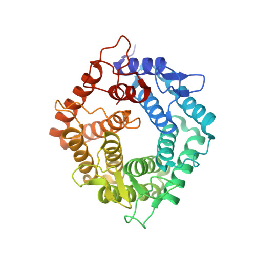 | |
UniProt | |||||
Find proteins for Q9Z4P9 (Niallia circulans) Explore Q9Z4P9 Go to UniProtKB: Q9Z4P9 | |||||
Entity Groups | |||||
| Sequence Clusters | 30% Identity50% Identity70% Identity90% Identity95% Identity100% Identity | ||||
| UniProt Group | Q9Z4P9 | ||||
Sequence AnnotationsExpand | |||||
| |||||
| Length ( Å ) | Angle ( ˚ ) |
|---|---|
| a = 50.948 | α = 90 |
| b = 65.093 | β = 90 |
| c = 90.242 | γ = 90 |
| Software Name | Purpose |
|---|---|
| PHENIX | refinement |
RCSB PDB (citation) is hosted by
RCSB PDB is a member of the

