4BJH

Crystal Structure of the Aquifex Reactor Complex Formed by Dihydroorotase (H180A, H232A) with Dihydroorotate and Aspartate Transcarbamoylase with N-(phosphonacetyl)-L-aspartate (PALA)


Experimental Data Snapshot

  • Method: X-RAY DIFFRACTION
  • Resolution: 2.20 Å
  • R-Value Free: 0.203 
  • R-Value Work: 0.161 
  • R-Value Observed: 0.163 

wwPDB Validation   3D Report Full Report


Ligand Structure Quality Assessment 


This is version 1.3 of the entry. See complete history


Literature

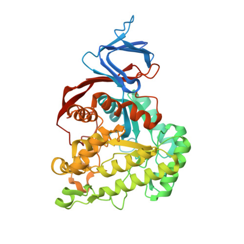
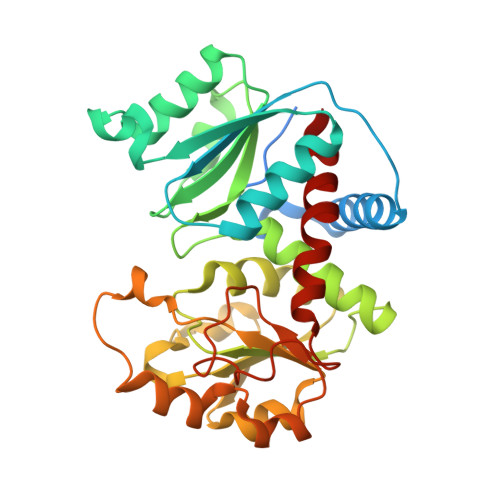
The Mononuclear Metal Center of Type-I Dihydroorotase from Aquifex Aeolicus.

Edwards, B.F.Fernando, R.Martin, P.D.Grimley, E.Cordes, M.Vaishnav, A.Brunzelle, J.S.Evans, H.G.Evans, D.R.

(2013) BMC Biochem 14: 36

  • DOI: https://doi.org/10.1186/1471-2091-14-36
  • Primary Citation of Related Structures:  
    4BJH

  • PubMed Abstract: 

    Dihydroorotase (DHO) is a zinc metalloenzyme, although the number of active site zinc ions has been controversial. E. coli DHO was initially thought to have a mononuclear metal center, but the subsequent X-ray structure clearly showed two zinc ions, α and β, at the catalytic site. Aquifex aeolicus DHO, is a dodecamer comprised of six DHO and six aspartate transcarbamoylase (ATC) subunits. The isolated DHO monomer, which lacks catalytic activity, has an intact α-site and conserved β-site ligands, but the geometry of the second metal binding site is completely disrupted. However, the putative β-site is restored when the complex with ATC is formed and DHO activity is regained. Nevertheless, the X-ray structure of the complex revealed a single zinc ion at the active site. The structure of DHO from the pathogenic organism, S. aureus showed that it also has a single active site metal ion. Zinc analysis showed that the enzyme has one zinc/DHO subunit and the addition of excess metal ion did not stimulate catalytic activity, nor alter the kinetic parameters. The metal free apoenzyme was inactive, but the full activity was restored upon the addition of one equivalent of Zn2+ or Co2+. Moreover, deletion of the β-site by replacing the His180 and His232 with alanine had no effect on catalysis in the presence or absence of excess zinc. The 2.2 Å structure of the double mutant confirmed that the β-site was eliminated but that the active site remained otherwise intact. Thus, kinetically competent A. aeolicus DHO has a mononuclear metal center. In contrast, elimination of the putative second metal binding site in amidohydrolyases with a binuclear metal center, resulted in the abolition of catalytic activity. The number of active site metal ions may be a consideration in the design of inhibitors that selectively target either the mononuclear or binuclear enzymes.


  • Organizational Affiliation

    Department of Biochemistry and Molecular Biology, Wayne State University School of Medicine, 540 East Canfield Street, Detroit, MI 48201, USA. drevans@med.wayne.edu.


Macromolecules
Find similar proteins by:  (by identity cutoff)  |  3D Structure
Entity ID: 1
MoleculeChains Sequence LengthOrganismDetailsImage
DIHYDROOROTASE456Aquifex aeolicusMutation(s): 2 
EC: 3.5.2.3
UniProt
Find proteins for O66990 (Aquifex aeolicus (strain VF5))
Explore O66990 
Go to UniProtKB:  O66990
Entity Groups  
Sequence Clusters30% Identity50% Identity70% Identity90% Identity95% Identity100% Identity
UniProt GroupO66990
Sequence Annotations
Expand
  • Reference Sequence
Find similar proteins by:  (by identity cutoff)  |  3D Structure
Entity ID: 2
MoleculeChains Sequence LengthOrganismDetailsImage
ASPARTATE CARBAMOYLTRANSFERASE322Aquifex aeolicusMutation(s): 0 
EC: 2.1.3.2
UniProt
Find proteins for O66726 (Aquifex aeolicus (strain VF5))
Explore O66726 
Go to UniProtKB:  O66726
Entity Groups  
Sequence Clusters30% Identity50% Identity70% Identity90% Identity95% Identity100% Identity
UniProt GroupO66726
Sequence Annotations
Expand
  • Reference Sequence
Small Molecules
Ligands 6 Unique
IDChains Name / Formula / InChI Key2D Diagram3D Interactions
PAL
Query on PAL

Download Ideal Coordinates CCD File 
E [auth B]N-(PHOSPHONACETYL)-L-ASPARTIC ACID
C6 H10 N O8 P
ZZKNRXZVGOYGJT-VKHMYHEASA-N
DOR
Query on DOR

Download Ideal Coordinates CCD File 
D [auth A](4S)-2,6-DIOXOHEXAHYDROPYRIMIDINE-4-CARBOXYLIC ACID
C5 H6 N2 O4
UFIVEPVSAGBUSI-REOHCLBHSA-N
BA
Query on BA

Download Ideal Coordinates CCD File 
G [auth B],
H [auth B],
I [auth B]
BARIUM ION
Ba
XDFCIPNJCBUZJN-UHFFFAOYSA-N
PO4
Query on PO4

Download Ideal Coordinates CCD File 
F [auth B]PHOSPHATE ION
O4 P
NBIIXXVUZAFLBC-UHFFFAOYSA-K
ZN
Query on ZN

Download Ideal Coordinates CCD File 
C [auth A]ZINC ION
Zn
PTFCDOFLOPIGGS-UHFFFAOYSA-N
EDO
Query on EDO

Download Ideal Coordinates CCD File 
J [auth B]
K [auth B]
L [auth B]
M [auth B]
N [auth B]
J [auth B],
K [auth B],
L [auth B],
M [auth B],
N [auth B],
O [auth B],
P [auth B],
Q [auth B],
R [auth B],
S [auth B],
T [auth B],
U [auth B]
1,2-ETHANEDIOL
C2 H6 O2
LYCAIKOWRPUZTN-UHFFFAOYSA-N
Experimental Data & Validation

Experimental Data

  • Method: X-RAY DIFFRACTION
  • Resolution: 2.20 Å
  • R-Value Free: 0.203 
  • R-Value Work: 0.161 
  • R-Value Observed: 0.163 
  • Space Group: H 3 2
Unit Cell:
Length ( Å )Angle ( ˚ )
a = 157.152α = 90
b = 157.152β = 90
c = 233.244γ = 120
Software Package:
Software NamePurpose
REFMACrefinement
MOSFLMdata reduction
SCALEPACKdata scaling
ARP/wARPphasing

Structure Validation

View Full Validation Report


Ligand Structure Quality Assessment 


Entry History 

Deposition Data

Revision History  (Full details and data files)

  • Version 1.0: 2013-12-18
    Type: Initial release
  • Version 1.1: 2017-06-28
    Changes: Refinement description
  • Version 1.2: 2019-05-29
    Changes: Data collection, Experimental preparation
  • Version 1.3: 2023-12-20
    Changes: Data collection, Database references, Derived calculations, Other, Refinement description