4BJ3

Integrin alpha2 I domain E318W-collagen complex


Experimental Data Snapshot

  • Method: X-RAY DIFFRACTION
  • Resolution: 3.04 Å
  • R-Value Free: 0.298 
  • R-Value Work: 0.246 
  • R-Value Observed: 0.251 

wwPDB Validation   3D Report Full Report


This is version 1.2 of the entry. See complete history


Literature

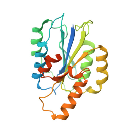
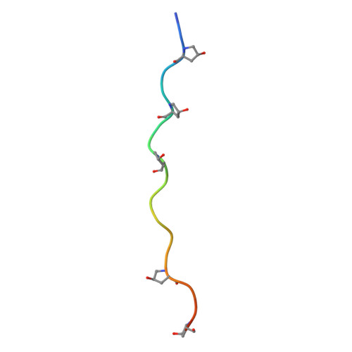
An Activating Mutation Reveals a Second Binding Mode of the Integrin Alpha2 I Domain to the Gfoger Motif in Collagens.

Carafoli, F.Hamaia, S.W.Bihan, D.Hohenester, E.Farndale, R.W.

(2013) PLoS One 8: 69833

  • DOI: https://doi.org/10.1371/journal.pone.0069833
  • Primary Citation of Related Structures:  
    4BJ3

  • PubMed Abstract: 

    The GFOGER motif in collagens (O denotes hydroxyproline) represents a high-affinity binding site for all collagen-binding integrins. Other GxOGER motifs require integrin activation for maximal binding. The E318W mutant of the integrin α2β1 I domain displays a relaxed collagen specificity, typical of an active state. E318W binds more strongly than the wild-type α2 I domain to GMOGER, and forms a 2:1 complex with a homotrimeric, collagen-like, GFOGER peptide. Crystal structure analysis of this complex reveals two E318W I domains, A and B, bound to a single triple helix. The E318W I domains are virtually identical to the collagen-bound wild-type I domain, suggesting that the E318W mutation activates the I domain by destabilising the unligated conformation. E318W I domain A interacts with two collagen chains similarly to wild-type I domain (high-affinity mode). E318W I domain B makes favourable interactions with only one collagen chain (low-affinity mode). This observation suggests that single GxOGER motifs in the heterotrimeric collagens V and IX may support binding of activated integrins.


  • Organizational Affiliation

    Department of Life Sciences, Imperial College London, London, United Kingdom.


Macromolecules
Find similar proteins by:  (by identity cutoff)  |  3D Structure
Entity ID: 1
MoleculeChains Sequence LengthOrganismDetailsImage
INTEGRIN ALPHA-2
A, B
225Homo sapiensMutation(s): 1 
UniProt & NIH Common Fund Data Resources
Find proteins for P17301 (Homo sapiens)
Explore P17301 
Go to UniProtKB:  P17301
PHAROS:  P17301
GTEx:  ENSG00000164171 
Entity Groups  
Sequence Clusters30% Identity50% Identity70% Identity90% Identity95% Identity100% Identity
UniProt GroupP17301
Sequence Annotations
Expand
  • Reference Sequence

Find similar proteins by:  Sequence   |   3D Structure  

Entity ID: 2
MoleculeChains Sequence LengthOrganismDetailsImage
GFOGER PEPTIDE
C, D, E
21Homo sapiensMutation(s): 0 
UniProt & NIH Common Fund Data Resources
Find proteins for Q96A83 (Homo sapiens)
Explore Q96A83 
Go to UniProtKB:  Q96A83
PHAROS:  Q96A83
GTEx:  ENSG00000160963 
Entity Groups  
Sequence Clusters30% Identity50% Identity70% Identity90% Identity95% Identity100% Identity
UniProt GroupQ96A83
Sequence Annotations
Expand
  • Reference Sequence
Small Molecules
Modified Residues  1 Unique
IDChains TypeFormula2D DiagramParent
HYP
Query on HYP
C, D, E
L-PEPTIDE LINKINGC5 H9 N O3PRO
Experimental Data & Validation

Experimental Data

  • Method: X-RAY DIFFRACTION
  • Resolution: 3.04 Å
  • R-Value Free: 0.298 
  • R-Value Work: 0.246 
  • R-Value Observed: 0.251 
  • Space Group: P 43 21 2
Unit Cell:
Length ( Å )Angle ( ˚ )
a = 83.004α = 90
b = 83.004β = 90
c = 175.256γ = 90
Software Package:
Software NamePurpose
PHENIXrefinement
XDSdata reduction
Aimlessdata scaling
PHASERphasing

Structure Validation

View Full Validation Report


Entry History 

Deposition Data

Revision History  (Full details and data files)

  • Version 1.0: 2013-11-20
    Type: Initial release
  • Version 1.1: 2015-10-21
    Changes: Source and taxonomy
  • Version 1.2: 2023-12-20
    Changes: Data collection, Database references, Derived calculations, Other, Refinement description