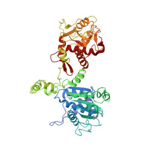
Large Terminase Conformational Change Induced by Connector Binding in Bacteriophage T7
Dauden, M.I., Martin-Benito, J., Sanchez-Ferrero, J.C., Pulido-Cid, M., Valpuesta, J.M., Carrascosa, J.L.(2013) J Biol Chem 288: 16998
- PubMed: 23632014
- DOI: https://doi.org/10.1074/jbc.M112.448951
- Primary Citation of Related Structures:
4BIJ, 4BIL - PubMed Abstract:
During bacteriophage morphogenesis DNA is translocated into a preformed prohead by the complex formed by the portal protein, or connector, plus the terminase, which are located at an especial prohead vertex. The terminase is a powerful motor that converts ATP hydrolysis into mechanical movement of the DNA. Here, we have determined the structure of the T7 large terminase by electron microscopy. The five terminase subunits assemble in a toroid that encloses a channel wide enough to accommodate dsDNA. The structure of the complete connector-terminase complex is also reported, revealing the coupling between the terminase and the connector forming a continuous channel. The structure of the terminase assembled into the complex showed a different conformation when compared with the isolated terminase pentamer. To understand in molecular terms the terminase morphological change, we generated the terminase atomic model based on the crystallographic structure of its phage T4 counterpart. The docking of the threaded model in both terminase conformations showed that the transition between the two states can be achieved by rigid body subunit rotation in the pentameric assembly. The existence of two terminase conformations and its possible relation to the sequential DNA translocation may shed light into the molecular bases of the packaging mechanism of bacteriophage T7.
Organizational Affiliation:
Department of Macromolecular Structure, 28049 Madrid, Spain.