4BH5

LytM domain of EnvC, an activator of cell wall amidases in Escherichia coli


Experimental Data Snapshot

  • Method: X-RAY DIFFRACTION
  • Resolution: 1.57 Å
  • R-Value Free: 0.207 
  • R-Value Work: 0.166 
  • R-Value Observed: 0.170 

wwPDB Validation   3D Report Full Report


This is version 1.3 of the entry. See complete history


Literature

Structure-Function Analysis of the Lytm Domain of Envc, an Activator of Cell Wall Remodeling at the Escherichia Coli Division Site.

Peters, N.T.Morlot, C.Yang, D.C.Uehara, T.Vernet, T.Bernhardt, T.G.

(2013) Mol Microbiol 89: 690

  • DOI: https://doi.org/10.1111/mmi.12304
  • Primary Citation of Related Structures:  
    4BH5

  • PubMed Abstract: 

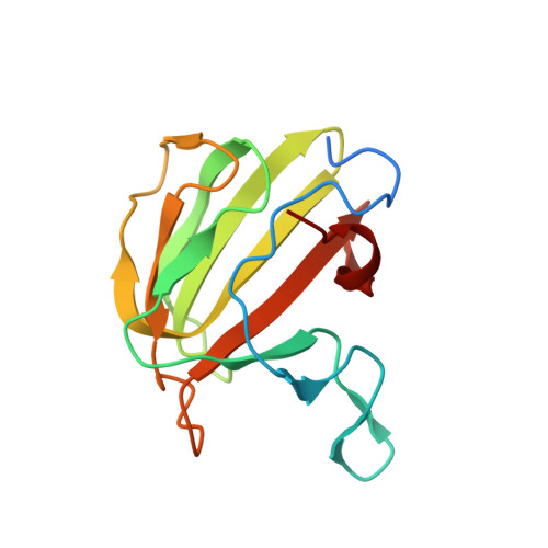
    Proteins with LytM (Peptidase_M23) domains are broadly distributed in bacteria and have been implicated in a variety of important processes, including cell division and cell-shape determination. Most LytM-like proteins that have been structurally and/or biochemically characterized are metallo-endopeptidases that cleave cross-links in the peptidoglycan (PG) cell wall matrix. Notable exceptions are the Escherichia coli cell division proteins EnvC and NlpD. These LytM factors are not hydrolases themselves, but instead serve as activators that stimulate PG cleavage by target enzymes called amidases to promote cell separation. Here we report the structure of the LytM domain from EnvC, the first structure of a LytM factor implicated in the regulation of PG hydrolysis. As expected, the fold is highly similar to that of other LytM proteins. However, consistent with its role as a regulator, the active-site region is degenerate and lacks a catalytic metal ion. Importantly, genetic analysis indicates that residues in and around this degenerate active site are critical for amidase activation in vivo and in vitro. Thus, in the regulatory LytM factors, the apparent substrate binding pocket conserved in active metallo-endopeptidases has been adapted to control PG hydrolysis by another set of enzymes.


  • Organizational Affiliation

    Department of Microbiology and Immunobiology, Harvard Medical School, 200 Longwood Avenue, Boston, MA 02115, USA.


Macromolecules
Find similar proteins by:  (by identity cutoff)  |  3D Structure
Entity ID: 1
MoleculeChains Sequence LengthOrganismDetailsImage
MUREIN HYDROLASE ACTIVATOR ENVC
A, B, C, D
142Escherichia coli K-12Mutation(s): 0 
UniProt
Find proteins for P37690 (Escherichia coli (strain K12))
Explore P37690 
Go to UniProtKB:  P37690
Entity Groups  
Sequence Clusters30% Identity50% Identity70% Identity90% Identity95% Identity100% Identity
UniProt GroupP37690
Sequence Annotations
Expand
  • Reference Sequence
Small Molecules
Ligands 4 Unique
IDChains Name / Formula / InChI Key2D Diagram3D Interactions
IOD
Query on IOD

Download Ideal Coordinates CCD File 
BA [auth D]
CA [auth D]
DA [auth D]
E [auth A]
EA [auth D]
BA [auth D],
CA [auth D],
DA [auth D],
E [auth A],
EA [auth D],
F [auth A],
G [auth A],
H [auth A],
HA [auth D],
J [auth B],
K [auth B],
L [auth B],
M [auth B],
O [auth B],
P [auth B],
Q [auth B],
R [auth B],
S [auth C],
T [auth C],
U [auth C],
V [auth C],
W [auth C],
Z [auth C]
IODIDE ION
I
XMBWDFGMSWQBCA-UHFFFAOYSA-M
GOL
Query on GOL

Download Ideal Coordinates CCD File 
GA [auth D],
Y [auth C]
GLYCEROL
C3 H8 O3
PEDCQBHIVMGVHV-UHFFFAOYSA-N
K
Query on K

Download Ideal Coordinates CCD File 
AA [auth C]POTASSIUM ION
K
NPYPAHLBTDXSSS-UHFFFAOYSA-N
CL
Query on CL

Download Ideal Coordinates CCD File 
FA [auth D],
I [auth A],
N [auth B],
X [auth C]
CHLORIDE ION
Cl
VEXZGXHMUGYJMC-UHFFFAOYSA-M
Experimental Data & Validation

Experimental Data

  • Method: X-RAY DIFFRACTION
  • Resolution: 1.57 Å
  • R-Value Free: 0.207 
  • R-Value Work: 0.166 
  • R-Value Observed: 0.170 
  • Space Group: P 31
Unit Cell:
Length ( Å )Angle ( ˚ )
a = 57.404α = 90
b = 57.404β = 90
c = 129.32γ = 120
Software Package:
Software NamePurpose
REFMACrefinement
XDSdata reduction
XSCALEdata scaling
PHASERphasing

Structure Validation

View Full Validation Report


Entry History 

Deposition Data

Revision History  (Full details and data files)

  • Version 1.0: 2013-07-03
    Type: Initial release
  • Version 1.1: 2013-08-28
    Changes: Database references
  • Version 1.2: 2019-10-16
    Changes: Data collection, Experimental preparation, Other
  • Version 1.3: 2023-12-20
    Changes: Data collection, Database references, Derived calculations, Refinement description