4BGD

Crystal structure of Brr2 in complex with the Jab1/MPN domain of Prp8


Experimental Data Snapshot

  • Method: X-RAY DIFFRACTION
  • Resolution: 3.10 Å
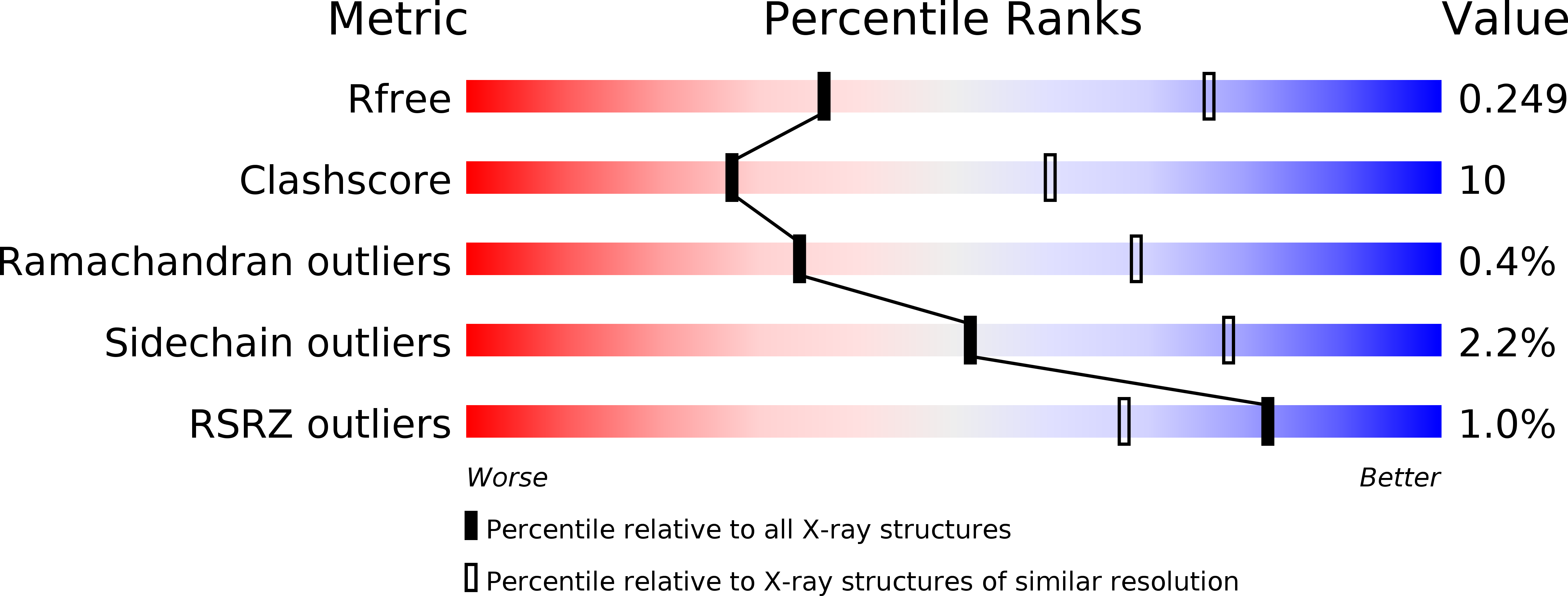
  • R-Value Free: 0.270 
  • R-Value Work: 0.214 
  • R-Value Observed: 0.217 

wwPDB Validation   3D Report Full Report


Ligand Structure Quality Assessment 


This is version 1.3 of the entry. See complete history


Literature

Structural Basis of Brr2-Prp8 Interactions and Implications for U5 Snrnp Biogenesis and the Spliceosome Active Site

Nguyen, T.H.D.Li, J.Galej, W.P.Oshikane, H.Newman, A.J.Nagai, K.

(2013) Structure 21: 910

  • DOI: https://doi.org/10.1016/j.str.2013.04.017
  • Primary Citation of Related Structures:  
    4BGD

  • PubMed Abstract: 

    The U5 small nuclear ribonucleoprotein particle (snRNP) helicase Brr2 disrupts the U4/U6 small nuclear RNA (snRNA) duplex and allows U6 snRNA to engage in an intricate RNA network at the active center of the spliceosome. Here, we present the structure of yeast Brr2 in complex with the Jab1/MPN domain of Prp8, which stimulates Brr2 activity. Contrary to previous reports, our crystal structure and mutagenesis data show that the Jab1/MPN domain binds exclusively to the N-terminal helicase cassette. The residues in the Jab1/MPN domain, whose mutations in human Prp8 cause the degenerative eye disease retinitis pigmentosa, are found at or near the interface with Brr2, clarifying its molecular pathology. In the cytoplasm, Prp8 forms a precursor complex with U5 snRNA, seven Smproteins, Snu114, and Aar2, but after nuclear import, Brr2 replaces Aar2 to form mature U5 snRNP. Our structure explains why Aar2 and Brr2 are mutually exclusive and provides important insights into the assembly of U5 snRNP.


  • Organizational Affiliation

    MRC Laboratory of Molecular Biology, Francis Crick Avenue, Cambridge CB2 0QH, UK.


Macromolecules
Find similar proteins by:  (by identity cutoff)  |  3D Structure
Entity ID: 1
MoleculeChains Sequence LengthOrganismDetailsImage
PRE-MRNA-SPLICING HELICASE BRR21,722Saccharomyces cerevisiaeMutation(s): 0 
EC: 3.6.4.13
UniProt
Find proteins for P32639 (Saccharomyces cerevisiae (strain ATCC 204508 / S288c))
Explore P32639 
Go to UniProtKB:  P32639
Entity Groups  
Sequence Clusters30% Identity50% Identity70% Identity90% Identity95% Identity100% Identity
UniProt GroupP32639
Sequence Annotations
Expand
  • Reference Sequence
Find similar proteins by:  (by identity cutoff)  |  3D Structure
Entity ID: 2
MoleculeChains Sequence LengthOrganismDetailsImage
PRE-MRNA-SPLICING FACTOR 8B [auth C]248Saccharomyces cerevisiaeMutation(s): 0 
UniProt
Find proteins for P33334 (Saccharomyces cerevisiae (strain ATCC 204508 / S288c))
Explore P33334 
Go to UniProtKB:  P33334
Entity Groups  
Sequence Clusters30% Identity50% Identity70% Identity90% Identity95% Identity100% Identity
UniProt GroupP33334
Sequence Annotations
Expand
  • Reference Sequence
Small Molecules
Experimental Data & Validation

Experimental Data

  • Method: X-RAY DIFFRACTION
  • Resolution: 3.10 Å
  • R-Value Free: 0.270 
  • R-Value Work: 0.214 
  • R-Value Observed: 0.217 
  • Space Group: P 21 21 21
Unit Cell:
Length ( Å )Angle ( ˚ )
a = 107.64α = 90
b = 178.64β = 90
c = 180.4γ = 90
Software Package:
Software NamePurpose
REFMACrefinement
iMOSFLMdata reduction
Aimlessdata scaling
SHARPphasing

Structure Validation

View Full Validation Report


Ligand Structure Quality Assessment 


Entry History 

Deposition Data

Revision History  (Full details and data files)

  • Version 1.0: 2013-05-29
    Type: Initial release
  • Version 1.1: 2013-06-12
    Changes: Database references
  • Version 1.2: 2013-08-14
    Changes: Database references
  • Version 1.3: 2023-12-20
    Changes: Data collection, Database references, Derived calculations, Other, Refinement description, Structure summary