4BAE

Optimisation of pyrroleamides as mycobacterial GyrB ATPase inhibitors: Structure Activity Relationship and in vivo efficacy in the mouse model of tuberculosis


Experimental Data Snapshot

  • Method: X-RAY DIFFRACTION
  • Resolution: 2.35 Å
  • R-Value Free: 0.237 
  • R-Value Work: 0.194 
  • R-Value Observed: 0.196 

wwPDB Validation   3D Report Full Report


Ligand Structure Quality Assessment 


This is version 1.4 of the entry. See complete history


Literature

Optimization of Pyrrolamides as Mycobacterial Gyrb ATPase Inhibitors: Structure Activity Relationship and in Vivo Efficacy in the Mouse Model of Tuberculosis.

Hameed, S.P.Solapure, S.Mukherjee, K.Nandi, V.Waterson, D.Shandil, R.Balganesh, M.Sambandamurthy, V.K.Raichurkar, A.K.Deshpande, A.Ghosh, A.Awasthy, D.Shanbhag, G.Sheikh, G.Mcmiken, H.Puttur, J.Reddy, J.Werngren, J.Read, J.Kumar, M.Manjunatha, R.Chinnapattu, M.Madhavapeddi, P.Manjrekar, P.Basu, R.Gaonkar, S.Sharma, S.Hoffner, S.Humnabadkar, V.Subbulakshmi, V.Panduga, V.

(2014) Antimicrob Agents Chemother 58: 61

  • DOI: https://doi.org/10.1128/AAC.01751-13
  • Primary Citation of Related Structures:  
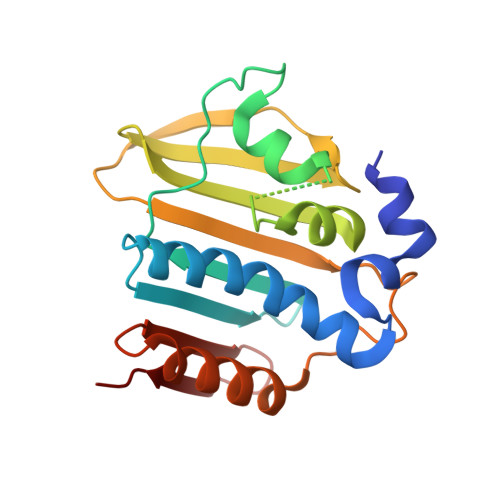
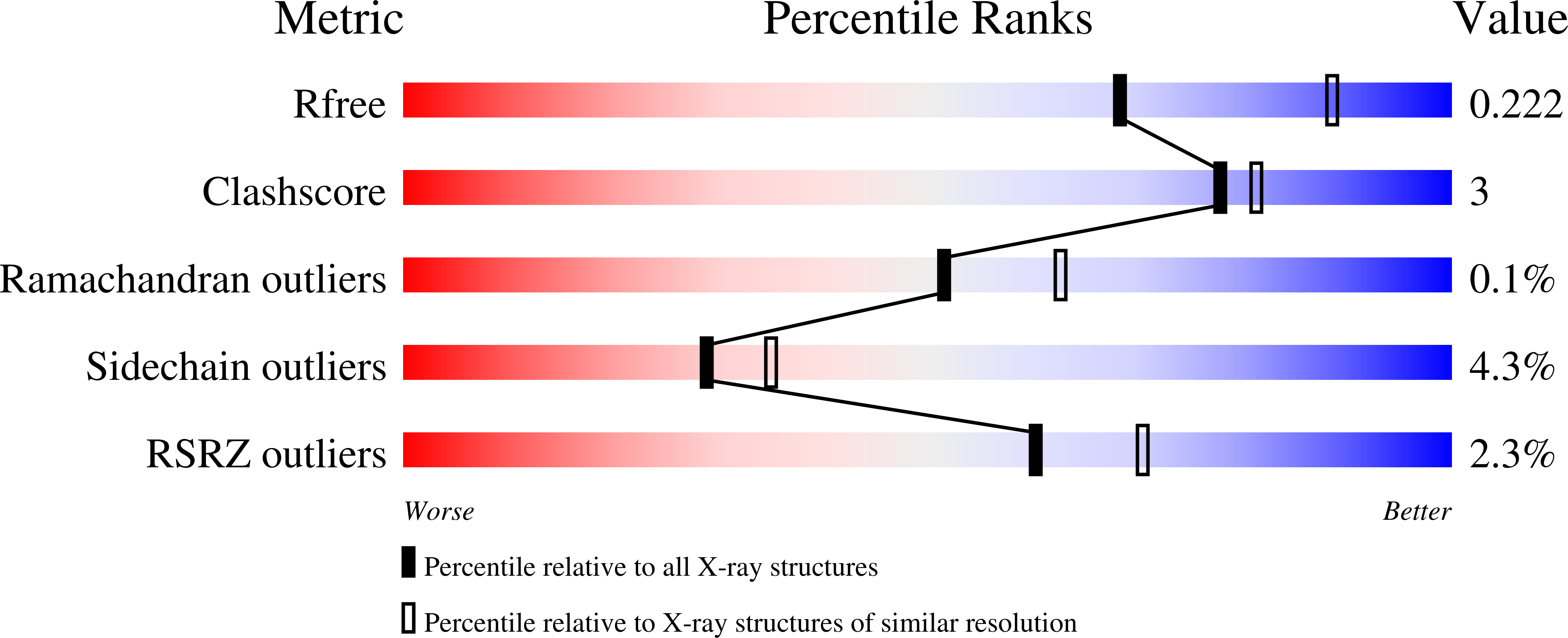
    4BAE

  • PubMed Abstract: 

    Moxifloxacin has shown excellent activity against drug-sensitive as well as drug-resistant tuberculosis (TB), thus confirming DNA gyrase as a clinically validated target for discovering novel anti-TB agents. We have identified novel inhibitors in the pyrrolamide class which kill Mycobacterium tuberculosis through inhibition of ATPase activity catalyzed by the GyrB domain of DNA gyrase. A homology model of the M. tuberculosis H37Rv GyrB domain was used for deciphering the structure-activity relationship and binding interactions of inhibitors with mycobacterial GyrB enzyme. Proposed binding interactions were later confirmed through cocrystal structure studies with the Mycobacterium smegmatis GyrB ATPase domain. The most potent compound in this series inhibited supercoiling activity of DNA gyrase with a 50% inhibitory concentration (IC50) of <5 nM, an MIC of 0.03 μg/ml against M. tuberculosis H37Rv, and an MIC90 of <0.25 μg/ml against 99 drug-resistant clinical isolates of M. tuberculosis. The frequency of isolating spontaneous resistant mutants was ∼10(-6) to 10(-8), and the point mutation mapped to the M. tuberculosis GyrB domain (Ser208 Ala), thus confirming its mode of action. The best compound tested for in vivo efficacy in the mouse model showed a 1.1-log reduction in lung CFU in the acute model and a 0.7-log reduction in the chronic model. This class of GyrB inhibitors could be developed as novel anti-TB agents.


  • Organizational Affiliation

    AstraZeneca India Pvt Ltd., Hebbal, Bangalore, India.


Macromolecules
Find similar proteins by:  (by identity cutoff)  |  3D Structure
Entity ID: 1
MoleculeChains Sequence LengthOrganismDetailsImage
DNA GYRASE SUBUNIT B
A, B, C, D
205Mycolicibacterium smegmatisMutation(s): 1 
EC: 5.99.1.3
UniProt
Find proteins for P0C559 (Mycolicibacterium smegmatis)
Explore P0C559 
Go to UniProtKB:  P0C559
Entity Groups  
Sequence Clusters30% Identity50% Identity70% Identity90% Identity95% Identity100% Identity
UniProt GroupP0C559
Sequence Annotations
Expand
  • Reference Sequence
Small Molecules
Ligands 3 Unique
IDChains Name / Formula / InChI Key2D Diagram3D Interactions
RWX
Query on RWX

Download Ideal Coordinates CCD File 
F [auth A],
H [auth B],
K [auth C],
N [auth D]
2-[(3S,4R)-4-[(3-bromanyl-4-chloranyl-5-methyl-1H-pyrrol-2-yl)carbonylamino]-3-methoxy-piperidin-1-yl]-4-(2-methyl-1,2,4-triazol-3-yl)-1,3-thiazole-5-carboxylic acid
C19 H21 Br Cl N7 O4 S
IXOKPDRRYHAGRW-ZJUUUORDSA-N
SO4
Query on SO4

Download Ideal Coordinates CCD File 
G [auth A],
I [auth B],
L [auth C],
O [auth D]
SULFATE ION
O4 S
QAOWNCQODCNURD-UHFFFAOYSA-L
CA
Query on CA

Download Ideal Coordinates CCD File 
E [auth A],
J [auth C],
M [auth D],
P [auth D]
CALCIUM ION
Ca
BHPQYMZQTOCNFJ-UHFFFAOYSA-N
Binding Affinity Annotations 
IDSourceBinding Affinity
RWX Binding MOAD:  4BAE IC50: 3 (nM) from 1 assay(s)
Experimental Data & Validation

Experimental Data

  • Method: X-RAY DIFFRACTION
  • Resolution: 2.35 Å
  • R-Value Free: 0.237 
  • R-Value Work: 0.194 
  • R-Value Observed: 0.196 
  • Space Group: P 1 21 1
Unit Cell:
Length ( Å )Angle ( ˚ )
a = 59.191α = 90
b = 78.249β = 90.01
c = 89.75γ = 90
Software Package:
Software NamePurpose
BUSTERrefinement
MOSFLMdata reduction
SCALEPACKdata scaling
AMoREphasing

Structure Validation

View Full Validation Report


Ligand Structure Quality Assessment 


Entry History 

Deposition Data

Revision History  (Full details and data files)

  • Version 1.0: 2013-10-30
    Type: Initial release
  • Version 1.1: 2014-01-15
    Changes: Database references
  • Version 1.2: 2016-12-14
    Changes: Database references
  • Version 1.3: 2017-11-08
    Changes: Source and taxonomy
  • Version 1.4: 2023-12-20
    Changes: Data collection, Database references, Derived calculations, Other, Refinement description