4B53

Crystal structure of the isolated IgG4 CH3 domain


Experimental Data Snapshot

  • Method: X-RAY DIFFRACTION
  • Resolution: 1.80 Å
  • R-Value Free: 0.237 
  • R-Value Work: 0.183 
  • R-Value Observed: 0.185 

wwPDB Validation   3D Report Full Report


This is version 2.1 of the entry. See complete history


Literature

Crystal Structure of the Human Igg4 C(H)3 Dimer Reveals the Role of Arg409 in the Mechanism of Fab-Arm Exchange.

Davies, A.M.Rispens, T.Den Bleker, T.H.Mcdonnell, J.M.Gould, H.J.Aalberse, R.C.Sutton, B.J.

(2012) Mol Immunol 54: 1

  • DOI: https://doi.org/10.1016/j.molimm.2012.10.029
  • Primary Citation of Related Structures:  
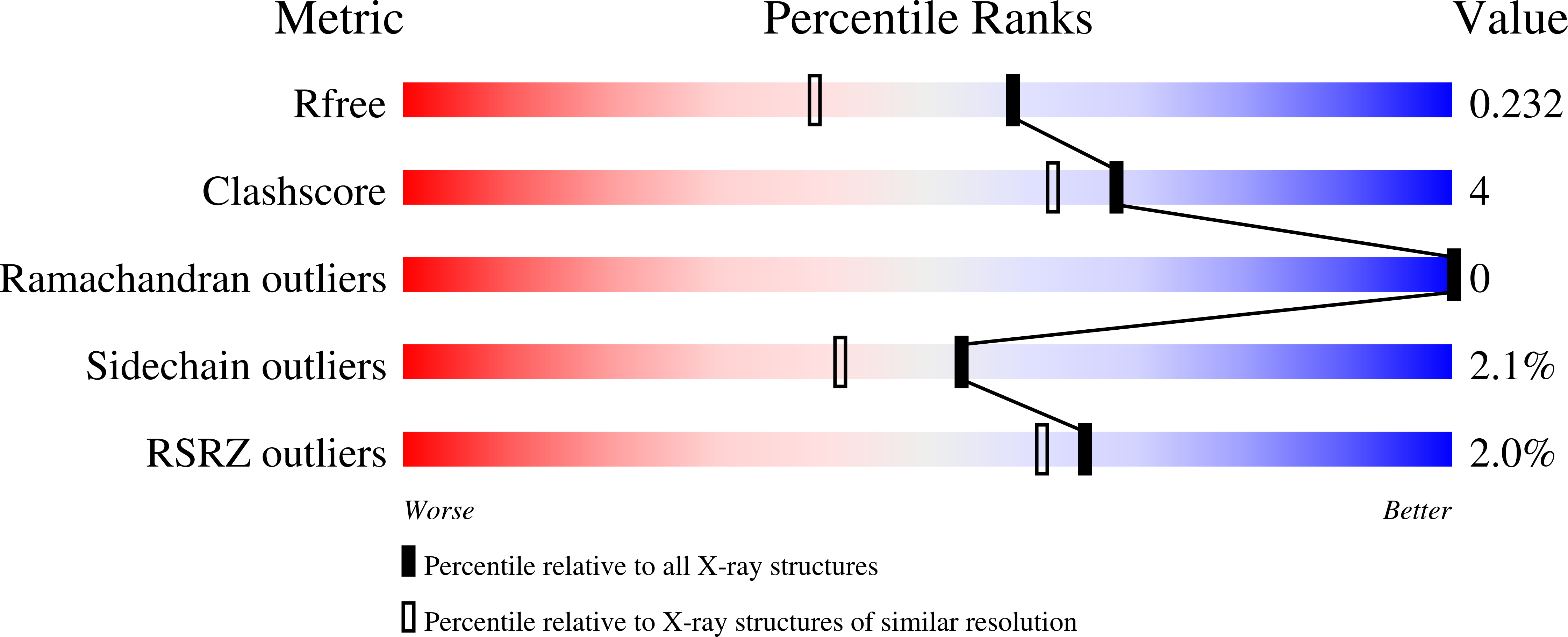
    4B53

  • PubMed Abstract: 

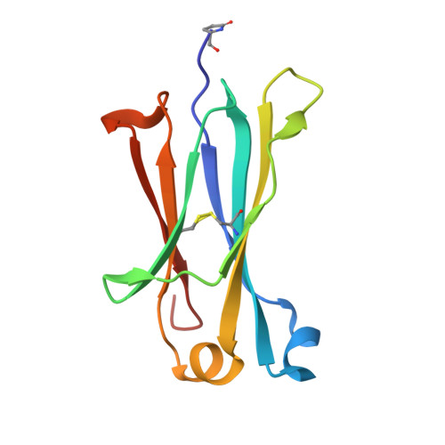
    Antibodies of the human IgG4 subclass uniquely undergo a process of Fab-arm exchange in which the heavy-chains of antibodies of different specificities can dissociate and then recombine. The mechanism by which the resulting functionally monovalent but bi-specific antibodies are formed is not only key to understanding their biological role, but is also important for the design of therapeutic monoclonal antibodies. Both the hinge region and the C(H)3 domain interface are known to be involved, and of the residues that differ between human IgG1 and IgG4 in C(H)3, residue 409, the only difference at the interface itself, has been implicated. We report the high resolution (1.8Å) structure of the C(H)3 domain dimer of IgG4, and find that Arg409 in IgG4, when compared with Lys409 observed in high resolution IgG1 structures, disrupts a network of water-mediated hydrogen bonding that is conserved in IgG1. Other conformational differences were detected that are a consequence of the presence of Arg409, such as a widening of the separation between residues Asn390 in one domain and Ser 400 in the other, which opens up a groove at the edge of the interface in IgG4 compared with IgG1. The effect of all these differences on the C(H)3 interface, doubled as a result of the interface's two-fold symmetry, is weakening of the inter-domain interaction in IgG4 compared with IgG1. This suggests a mechanism by which Arg409 weakens the C(H)3 interface in IgG4, predisposing this human antibody subclass to Fab-arm exchange.


  • Organizational Affiliation

    King's College London, Randall Division of Cell and Molecular Biophysics, London, SE1 1UL, United Kingdom.


Macromolecules
Find similar proteins by:  (by identity cutoff)  |  3D Structure
Entity ID: 1
MoleculeChains Sequence LengthOrganismDetailsImage
IG GAMMA-4 CHAIN C REGION
A, B
106Homo sapiensMutation(s): 0 
UniProt & NIH Common Fund Data Resources
Find proteins for P01861 (Homo sapiens)
Explore P01861 
Go to UniProtKB:  P01861
PHAROS:  P01861
GTEx:  ENSG00000211892 
Entity Groups  
Sequence Clusters30% Identity50% Identity70% Identity90% Identity95% Identity100% Identity
UniProt GroupP01861
Sequence Annotations
Expand
  • Reference Sequence
Small Molecules
Ligands 4 Unique
IDChains Name / Formula / InChI Key2D Diagram3D Interactions
TRS
Query on TRS

Download Ideal Coordinates CCD File 
H [auth A]2-AMINO-2-HYDROXYMETHYL-PROPANE-1,3-DIOL
C4 H12 N O3
LENZDBCJOHFCAS-UHFFFAOYSA-O
PEG
Query on PEG

Download Ideal Coordinates CCD File 
F [auth A],
G [auth A],
I [auth A],
M [auth B]
DI(HYDROXYETHYL)ETHER
C4 H10 O3
MTHSVFCYNBDYFN-UHFFFAOYSA-N
EDO
Query on EDO

Download Ideal Coordinates CCD File 
C [auth A],
D [auth A],
E [auth A],
K [auth B],
L [auth B]
1,2-ETHANEDIOL
C2 H6 O2
LYCAIKOWRPUZTN-UHFFFAOYSA-N
ACT
Query on ACT

Download Ideal Coordinates CCD File 
J [auth B]ACETATE ION
C2 H3 O2
QTBSBXVTEAMEQO-UHFFFAOYSA-M
Modified Residues  1 Unique
IDChains TypeFormula2D DiagramParent
PCA
Query on PCA
A, B
L-PEPTIDE LINKINGC5 H7 N O3GLN
Experimental Data & Validation

Experimental Data

  • Method: X-RAY DIFFRACTION
  • Resolution: 1.80 Å
  • R-Value Free: 0.237 
  • R-Value Work: 0.183 
  • R-Value Observed: 0.185 
  • Space Group: P 43 2 2
Unit Cell:
Length ( Å )Angle ( ˚ )
a = 57.09α = 90
b = 57.09β = 90
c = 138.436γ = 90
Software Package:
Software NamePurpose
PHENIXrefinement
XDSdata reduction
SCALAdata scaling
MOLREPphasing

Structure Validation

View Full Validation Report


Entry History 

Deposition Data

Revision History  (Full details and data files)

  • Version 1.0: 2012-12-05
    Type: Initial release
  • Version 1.1: 2013-07-31
    Changes: Derived calculations, Other
  • Version 1.2: 2013-09-04
    Changes: Atomic model, Derived calculations, Non-polymer description
  • Version 1.3: 2019-05-08
    Changes: Data collection, Derived calculations, Experimental preparation, Other
  • Version 1.4: 2019-05-22
    Changes: Data collection, Experimental preparation
  • Version 2.0: 2020-03-04
    Changes: Derived calculations, Other, Polymer sequence
  • Version 2.1: 2023-12-20
    Changes: Data collection, Database references, Derived calculations, Refinement description