4B4P

Crystal Structure of the lectin domain of F18 fimbrial adhesin FedF.


Experimental Data Snapshot

  • Method: X-RAY DIFFRACTION
  • Resolution: 1.80 Å
  • R-Value Free: 0.221 
  • R-Value Work: 0.178 
  • R-Value Observed: 0.179 

wwPDB Validation   3D Report Full Report


This is version 1.3 of the entry. See complete history

Re-refinement Note

A newer entry is available that reflects an alternative modeling of the original data: 4BWO


Literature

Structural Insight in Histo-Blood Group Binding by the F18 Fimbrial Adhesin Fedf.

Moonens, K.Bouckaert, J.Coddens, A.Tran, T.Panjikar, S.De Kerpel, M.Cox, E.Remaut, H.De Greve, H.

(2012) Mol Microbiol 86: 82

  • DOI: https://doi.org/10.1111/j.1365-2958.2012.08174.x
  • Primary Citation of Related Structures:  
    4B4P, 4B4Q, 4B4R

  • PubMed Abstract: 

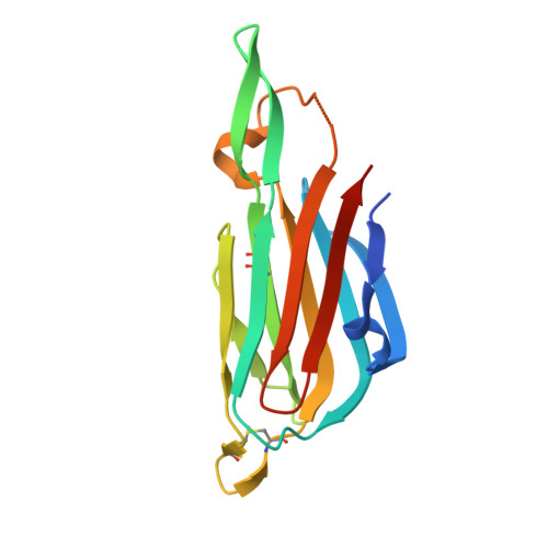
    F18-positive enterotoxigenic and Shiga toxin-producing Escherichia coli are responsible for post-weaning diarrhoea and oedema disease in pigs and lead to severe production losses in the farming industry. F18 fimbriae attach to the small intestine of young piglets by latching onto glycosphingolipids with A/H blood group determinants on type 1 core. We demonstrate the N-terminal domain of the F18 fimbrial subunit FedF to be responsible for ABH-mediated attachment and present its X-ray structure in ligand-free form and bound to A and B type 1 hexaoses. The FedF lectin domain comprises a 10-stranded immunoglobulin-like β-sandwich. Three linear motives, Q(47) -N(50), H(88) -S(90) and R(117) -T(119), form a shallow glycan binding pocket near the tip of the domain that is selective for type 1 core glycans in extended conformation. In addition to the glycan binding pocket, a polybasic loop on the membrane proximal surface of FedF lectin domain is shown to be required for binding to piglet enterocytes. Although dispensable for ABH glycan recognition, the polybasic surface adds binding affinity in the context of the host cell membrane, a mechanism that is proposed to direct ABH-glycan binding to cell-bound glycosphingolipids and could allow bacteria to avoid clearance by secreted glycoproteins.


  • Organizational Affiliation

    Structural & Molecular Microbiology, VIB Department of Structural Biology, VIB, Brussels, Belgium.


Macromolecules
Find similar proteins by:  (by identity cutoff)  |  3D Structure
Entity ID: 1
MoleculeChains Sequence LengthOrganismDetailsImage
F18 FIMBRIAL ADHESIN AC
A, B
151Escherichia coli K-12Mutation(s): 0 
UniProt
Find proteins for Q47212 (Escherichia coli)
Explore Q47212 
Go to UniProtKB:  Q47212
Entity Groups  
Sequence Clusters30% Identity50% Identity70% Identity90% Identity95% Identity100% Identity
UniProt GroupQ47212
Sequence Annotations
Expand
  • Reference Sequence
Small Molecules
Ligands 2 Unique
IDChains Name / Formula / InChI Key2D Diagram3D Interactions
SO4
Query on SO4

Download Ideal Coordinates CCD File 
I [auth A],
Q [auth B]
SULFATE ION
O4 S
QAOWNCQODCNURD-UHFFFAOYSA-L
BR
Query on BR

Download Ideal Coordinates CCD File 
C [auth A]
D [auth A]
E [auth A]
F [auth A]
G [auth A]
C [auth A],
D [auth A],
E [auth A],
F [auth A],
G [auth A],
H [auth A],
J [auth A],
K [auth B],
L [auth B],
M [auth B],
N [auth B],
O [auth B],
P [auth B]
BROMIDE ION
Br
CPELXLSAUQHCOX-UHFFFAOYSA-M
Experimental Data & Validation

Experimental Data

  • Method: X-RAY DIFFRACTION
  • Resolution: 1.80 Å
  • R-Value Free: 0.221 
  • R-Value Work: 0.178 
  • R-Value Observed: 0.179 
  • Space Group: P 21 21 21
Unit Cell:
Length ( Å )Angle ( ˚ )
a = 36.181α = 90
b = 74.382β = 90
c = 98.857γ = 90
Software Package:
Software NamePurpose
SIGMAAmodel building
MLPHAREmodel building
XSCALEdata scaling
SHELXCDphasing
PHASERphasing
SIGMAAphasing
RESOLVEphasing
BP3phasing
ABSphasing
MLPHAREphasing
REFMACrefinement
SHELXEphasing

Structure Validation

View Full Validation Report


Entry History 

Deposition Data

Revision History  (Full details and data files)

  • Version 1.0: 2012-08-15
    Type: Initial release
  • Version 1.1: 2012-10-03
    Changes: Database references, Source and taxonomy
  • Version 1.2: 2019-07-17
    Changes: Data collection
  • Version 1.3: 2019-10-16
    Changes: Data collection, Experimental preparation, Other