4B4D

Crystal structure of FAD-containing ferredoxin-NADP reductase from Xanthomonas axonopodis pv. citri


Experimental Data Snapshot

  • Method: X-RAY DIFFRACTION
  • Resolution: 1.50 Å
  • R-Value Free: 0.230 
  • R-Value Work: 0.207 
  • R-Value Observed: 0.209 

wwPDB Validation   3D Report Full Report


Ligand Structure Quality Assessment 


This is version 1.2 of the entry. See complete history


Literature

Crystal Structure of Fad-Containing Ferredoxin- Nadp Reductase from Xanthomonas Axonopodis Pv. Citri

Tondo, M.L.Hurtado-Guerrero, R.Sanchez-Azqueta, A.Ceccarelli, E.A.Medina, M.Orellano, E.G.Martinez-Julvez, M.

(2013) Biomed Res Int 2013: 06572

  • DOI: https://doi.org/10.1155/2013/906572
  • Primary Citation of Related Structures:  
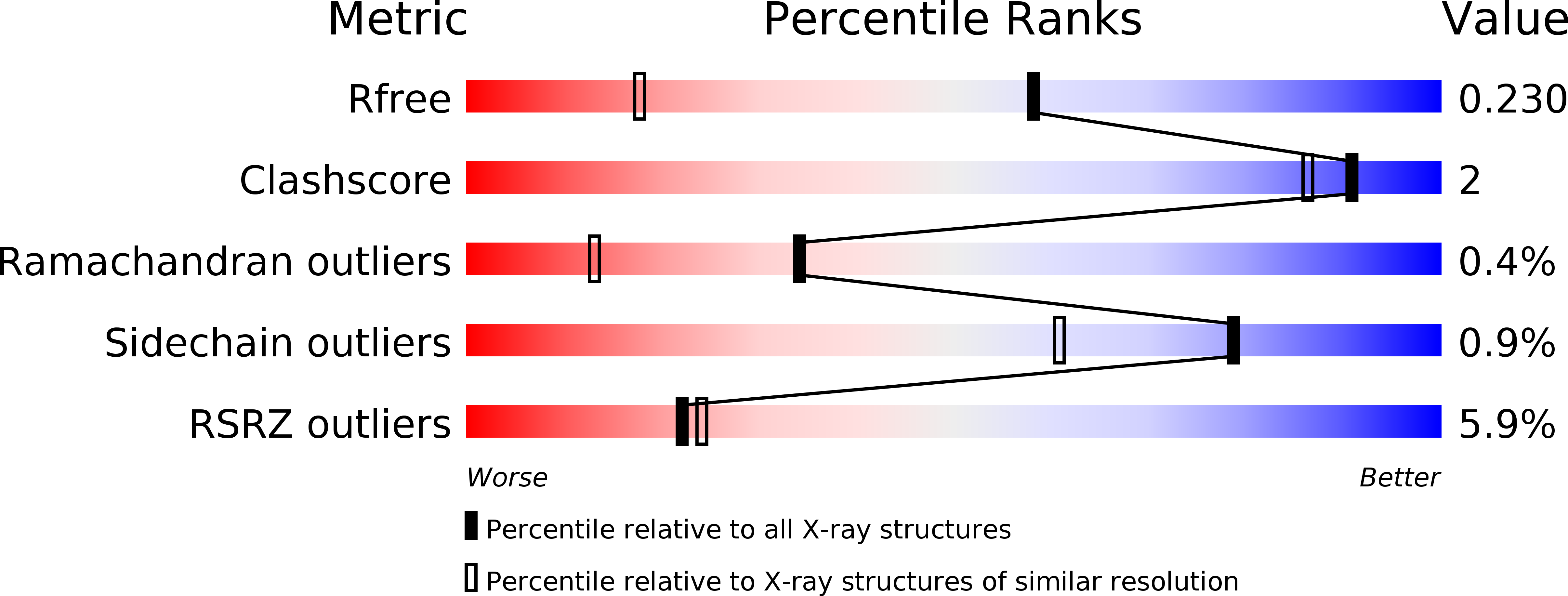
    4B4D

  • PubMed Abstract: 

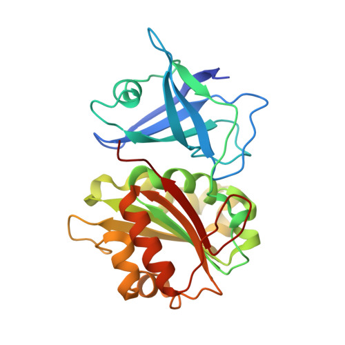
    We have solved the structure of ferredoxin-NADP(H) reductase, FPR, from the plant pathogen Xanthomonas axonopodis pv. citri, responsible for citrus canker, at a resolution of 1.5 Å. This structure reveals differences in the mobility of specific loops when compared to other FPRs, probably unrelated to the hydride transfer process, which contributes to explaining the structural and functional divergence between the subclass I FPRs. Interactions of the C-terminus of the enzyme with the phosphoadenosine of the cofactor FAD limit its mobility, thus affecting the entrance of nicotinamide into the active site. This structure opens the possibility of rationally designing drugs against the X. axonopodis pv. citri phytopathogen.


  • Organizational Affiliation

    Molecular Biology Division, Instituto de Biología Molecular y Celular de Rosario, IBR, CONICET, Facultad de Ciencias Bioquímicas y Farmacéuticas, Universidad Nacional de Rosario, Suipacha 531, S2002LRK Rosario, Argentina.


Macromolecules
Find similar proteins by:  (by identity cutoff)  |  3D Structure
Entity ID: 1
MoleculeChains Sequence LengthOrganismDetailsImage
FERREDOXIN-NADP REDUCTASE262Xanthomonas citri pv. citri str. 306Mutation(s): 0 
EC: 1.18.1.2
Entity Groups  
Sequence Clusters30% Identity50% Identity70% Identity90% Identity95% Identity100% Identity
Sequence Annotations
Expand
  • Reference Sequence
Small Molecules
Ligands 2 Unique
IDChains Name / Formula / InChI Key2D Diagram3D Interactions
FAD
Query on FAD

Download Ideal Coordinates CCD File 
B [auth A]FLAVIN-ADENINE DINUCLEOTIDE
C27 H33 N9 O15 P2
VWWQXMAJTJZDQX-UYBVJOGSSA-N
CL
Query on CL

Download Ideal Coordinates CCD File 
C [auth A]CHLORIDE ION
Cl
VEXZGXHMUGYJMC-UHFFFAOYSA-M
Experimental Data & Validation

Experimental Data

  • Method: X-RAY DIFFRACTION
  • Resolution: 1.50 Å
  • R-Value Free: 0.230 
  • R-Value Work: 0.207 
  • R-Value Observed: 0.209 
  • Space Group: I 2 2 2
Unit Cell:
Length ( Å )Angle ( ˚ )
a = 50.699α = 90
b = 99.775β = 90
c = 118.785γ = 90
Software Package:
Software NamePurpose
REFMACrefinement
XDSdata reduction
SCALEPACKdata scaling
MOLREPphasing

Structure Validation

View Full Validation Report


Ligand Structure Quality Assessment 


Entry History 

Deposition Data

Revision History  (Full details and data files)

  • Version 1.0: 2013-08-21
    Type: Initial release
  • Version 1.1: 2013-09-11
    Changes: Database references
  • Version 1.2: 2023-12-20
    Changes: Data collection, Database references, Derived calculations, Other, Refinement description