Structural and Functional Characterization of the Two Phosphoinositide Binding Sites of Proppins, a Beta-Propeller Protein Family.
Krick, R., Busse, R.A., Scacioc, A., Stephan, M., Janshoff, A., Thumm, M., Kuhnel, K.(2012) Proc Natl Acad Sci U S A 109: E2042
- PubMed: 22753491
- DOI: https://doi.org/10.1073/pnas.1205128109
- Primary Citation of Related Structures:
4AV8, 4AV9 - PubMed Abstract:
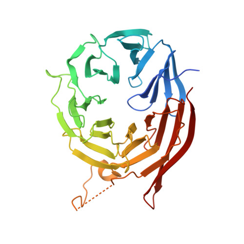
β-propellers that bind polyphosphoinositides (PROPPINs), a eukaryotic WD-40 motif-containing protein family, bind via their predicted β-propeller fold the polyphosphoinositides PtdIns3P and PtdIns(3,5)P(2) using a conserved FRRG motif. PROPPINs play a key role in macroautophagy in addition to other functions. We present the 3.0-Å crystal structure of Kluyveromyces lactis Hsv2, which shares significant sequence homologies with its three Saccharomyces cerevisiae homologs Atg18, Atg21, and Hsv2. It adopts a seven-bladed β-propeller fold with a rare nonvelcro propeller closure. Remarkably, in the crystal structure, the two arginines of the FRRG motif are part of two distinct basic pockets formed by a set of highly conserved residues. In comprehensive in vivo and in vitro studies of ScAtg18 and ScHsv2, we define within the two pockets a set of conserved residues essential for normal membrane association, phosphoinositide binding, and biological activities. Our experiments show that PROPPINs contain two individual phosphoinositide binding sites. Based on docking studies, we propose a model for phosphoinositide binding of PROPPINs.
Organizational Affiliation:
Department of Biochemistry II, Georg August University, D-37073 Göttingen, Germany.