4A5P

Structure of the Shigella flexneri MxiA protein


Experimental Data Snapshot

  • Method: X-RAY DIFFRACTION
  • Resolution: 3.15 Å
  • R-Value Free: 0.251 
  • R-Value Work: 0.231 
  • R-Value Observed: 0.232 

wwPDB Validation   3D Report Full Report


This is version 1.5 of the entry. See complete history


Literature

Architecture of the major component of the type III secretion system export apparatus.

Abrusci, P.Vergara-Irigaray, M.Johnson, S.Beeby, M.D.Hendrixson, D.R.Roversi, P.Friede, M.E.Deane, J.E.Jensen, G.J.Tang, C.M.Lea, S.M.

(2013) Nat Struct Mol Biol 20: 99-104

  • DOI: https://doi.org/10.1038/nsmb.2452
  • Primary Citation of Related Structures:  
    4A5P

  • PubMed Abstract: 

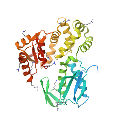
    Type III secretion systems (T3SSs) are bacterial membrane-embedded nanomachines designed to export specifically targeted proteins from the bacterial cytoplasm. Secretion through T3SS is governed by a subset of inner membrane proteins termed the 'export apparatus'. We show that a key member of the Shigella flexneri export apparatus, MxiA, assembles into a ring essential for secretion in vivo. The ring-forming interfaces are well-conserved in both nonflagellar and flagellar homologs, implying that the ring is an evolutionarily conserved feature in these systems. Electron cryo-tomography revealed a T3SS-associated cytoplasmic torus of size and shape corresponding to those of the MxiA ring aligned to the secretion channel located between the secretion pore and the ATPase complex. This defines the molecular architecture of the dominant component of the export apparatus and allows us to propose a model for the molecular mechanisms controlling secretion.


  • Organizational Affiliation

    Sir William Dunn School of Pathology, Oxford University, Oxford, UK.


Macromolecules
Find similar proteins by:  (by identity cutoff)  |  3D Structure
Entity ID: 1
MoleculeChains Sequence LengthOrganismDetailsImage
PROTEIN MXIA
A, B, C
383Shigella flexneri 5aMutation(s): 0 
UniProt
Find proteins for P0A1I5 (Shigella flexneri)
Explore P0A1I5 
Go to UniProtKB:  P0A1I5
Entity Groups  
Sequence Clusters30% Identity50% Identity70% Identity90% Identity95% Identity100% Identity
UniProt GroupP0A1I5
Sequence Annotations
Expand
  • Reference Sequence
Small Molecules
Modified Residues  1 Unique
IDChains TypeFormula2D DiagramParent
MLY
Query on MLY
A, B, C
L-PEPTIDE LINKINGC8 H18 N2 O2LYS
Experimental Data & Validation

Experimental Data

  • Method: X-RAY DIFFRACTION
  • Resolution: 3.15 Å
  • R-Value Free: 0.251 
  • R-Value Work: 0.231 
  • R-Value Observed: 0.232 
  • Space Group: P 63
Unit Cell:
Length ( Å )Angle ( ˚ )
a = 160.61α = 90
b = 160.61β = 90
c = 100.49γ = 120
Software Package:
Software NamePurpose
BUSTERrefinement
xia2data reduction
xia2data scaling
PHASERphasing

Structure Validation

View Full Validation Report


Entry History 

Deposition Data

Revision History  (Full details and data files)

  • Version 1.0: 2012-11-14
    Type: Initial release
  • Version 1.1: 2012-12-12
    Changes: Database references
  • Version 1.2: 2013-01-16
    Changes: Database references
  • Version 1.3: 2018-01-24
    Changes: Source and taxonomy
  • Version 1.4: 2019-05-08
    Changes: Data collection, Database references, Derived calculations, Experimental preparation
  • Version 1.5: 2023-12-20
    Changes: Data collection, Database references, Derived calculations, Other, Refinement description