4YWG

Crystal structure of 830A in complex with V1V2


Experimental Data Snapshot

  • Method: X-RAY DIFFRACTION
  • Resolution: 3.00 Å
  • R-Value Free: 0.296 
  • R-Value Work: 0.219 
  • R-Value Observed: 0.223 

wwPDB Validation   3D Report Full Report


Ligand Structure Quality Assessment 


This is version 1.6 of the entry. See complete history


Literature

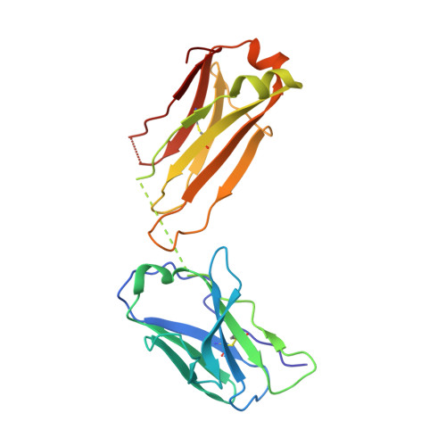
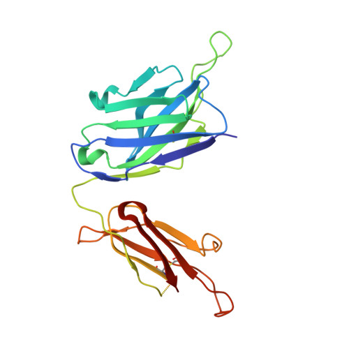
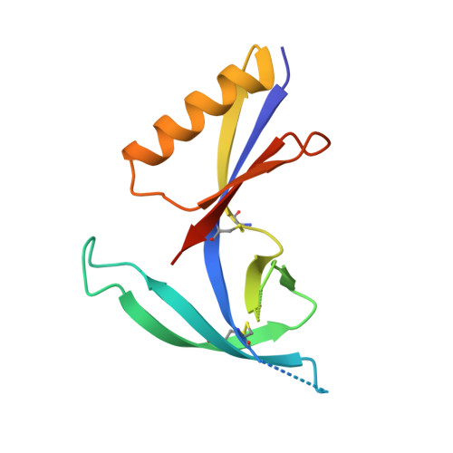
The V1V2 Region of HIV-1 gp120 Forms a Five-Stranded Beta Barrel.

Pan, R.Gorny, M.K.Zolla-Pazner, S.Kong, X.P.

(2015) J Virol 89: 8003-8010

  • DOI: https://doi.org/10.1128/JVI.00754-15
  • Primary Citation of Related Structures:  
    4YWG

  • PubMed Abstract: 

    The region consisting of the first and second variable regions (V1V2) of gp120 plays vital roles in the functioning of the HIV-1 envelope (Env). V1V2, which harbors multiple glycans and is highly sequence diverse, is located at the Env apex and stabilizes the trimeric gp120 spike on the virion surface. It shields V3 and the coreceptor binding sites in the prefusion state and exposes them upon CD4 binding. Data from the RV144 human HIV-1 vaccine trial suggested that antibody responses targeting the V1V2 region inversely correlated with the risk of infection; thus, understanding the antigenic structure of V1V2 can contribute to vaccine design. We have determined a crystal structure of a V1V2 scaffold molecule (V1V2ZM109-1FD6) in complex with 830A, a human monoclonal antibody that recognizes a V1V2 epitope overlapping the integrin-binding motif in V2. The structure revealed that V1V2 assumes a five-stranded beta barrel structure with the region of the integrin-binding site (amino acids [aa] 179 to 181) included in a "kink" followed by an extra beta strand. The complete barrel structure naturally presents the glycans on its outer surface and packs into its core conserved hydrophobic residues, including the Ile at position 181 which was highly correlated with vaccine efficacy in RV144. The epitope of monoclonal antibody 830A is discontinuous and composed of three segments: (i) Thr175, Tyr177, Leu179, and Asp180 at the kink overlapping the integrin-binding site; (ii) Arg153 and Val154 in V1; and (iii) Ile194 at the C terminus of V2. This report thus provides the atomic details of the immunogenic "V2i epitope."


  • Organizational Affiliation

    Departments of Biochemistry and Molecular Pharmacology, NYU School of Medicine, New York, New York, USA.


Macromolecules
Find similar proteins by:  (by identity cutoff)  |  3D Structure
Entity ID: 1
MoleculeChains Sequence LengthOrganismDetailsImage
Light chain of anti-HIV-1 gp120 V1V2 antibody 830AA [auth L],
D [auth M]
216Homo sapiensMutation(s): 0 
Entity Groups  
Sequence Clusters30% Identity50% Identity70% Identity90% Identity95% Identity100% Identity
Sequence Annotations
Expand
  • Reference Sequence
Find similar proteins by:  (by identity cutoff)  |  3D Structure
Entity ID: 2
MoleculeChains Sequence LengthOrganismDetailsImage
Heavy chain of anti-HIV-1 gp120 V1V2 antibody 830AB [auth H],
E [auth I]
226Homo sapiensMutation(s): 0 
Entity Groups  
Sequence Clusters30% Identity50% Identity70% Identity90% Identity95% Identity100% Identity
Sequence Annotations
Expand
  • Reference Sequence
Find similar proteins by:  (by identity cutoff)  |  3D Structure
Entity ID: 3
MoleculeChains Sequence LengthOrganismDetailsImage
Scaffold HIV-1 gp120 V1V2 region derived from strain ZM109C [auth G],
F [auth Q]
124Human immunodeficiency virus 1Mutation(s): 0 
UniProt
Find proteins for Q6TCP8 (Human immunodeficiency virus 1)
Explore Q6TCP8 
Go to UniProtKB:  Q6TCP8
Entity Groups  
Sequence Clusters30% Identity50% Identity70% Identity90% Identity95% Identity100% Identity
UniProt GroupQ6TCP8
Sequence Annotations
Expand
  • Reference Sequence
Experimental Data & Validation

Experimental Data

  • Method: X-RAY DIFFRACTION
  • Resolution: 3.00 Å
  • R-Value Free: 0.296 
  • R-Value Work: 0.219 
  • R-Value Observed: 0.223 
  • Space Group: P 21 21 21
Unit Cell:
Length ( Å )Angle ( ˚ )
a = 64.725α = 90
b = 159.168β = 90
c = 170.358γ = 90
Software Package:
Software NamePurpose
PHENIXrefinement
XDSdata reduction
PHENIXphasing
XDSdata scaling

Structure Validation

View Full Validation Report


Ligand Structure Quality Assessment 


Entry History & Funding Information

Deposition Data


Funding OrganizationLocationGrant Number
National Institutes of Health/National Institute Of Allergy and Infectious Diseases (NIH/NIAID)United StatesAI100151
National Institutes of Health/National Institute Of Allergy and Infectious Diseases (NIH/NIAID)United StatesAI082274
National Institutes of Health/National Institute Of Allergy and Infectious Diseases (NIH/NIAID)United StatesAI084119

Revision History  (Full details and data files)

  • Version 1.0: 2015-07-15
    Type: Initial release
  • Version 1.1: 2015-07-22
    Changes: Database references
  • Version 1.2: 2016-07-27
    Changes: Data collection
  • Version 1.3: 2017-09-13
    Changes: Author supporting evidence, Derived calculations
  • Version 1.4: 2019-12-11
    Changes: Author supporting evidence
  • Version 1.5: 2020-07-29
    Type: Remediation
    Reason: Carbohydrate remediation
    Changes: Data collection, Derived calculations, Structure summary
  • Version 1.6: 2023-09-27
    Changes: Data collection, Database references, Refinement description, Structure summary