4YRW

rat xanthine oxidoreductase, C-terminal deletion protein variant


Experimental Data Snapshot

  • Method: X-RAY DIFFRACTION
  • Resolution: 1.99 Å
  • R-Value Free: 0.229 
  • R-Value Work: 0.186 
  • R-Value Observed: 0.188 

wwPDB Validation   3D Report Full Report


Ligand Structure Quality Assessment 


This is version 1.3 of the entry. See complete history


Literature

The C-terminal peptide plays a role in the formation of an intermediate form during the transition between xanthine dehydrogenase and xanthine oxidase

Nishino, T.Okamoto, K.Kawaguchi, Y.Matsumura, T.Eger, B.T.Pai, E.F.Nishino, T.

(2015) FEBS J 282: 3075-3090

  • DOI: https://doi.org/10.1111/febs.13277
  • Primary Citation of Related Structures:  
    4YRW, 4YSW, 4YTY, 4YTZ

  • PubMed Abstract: 

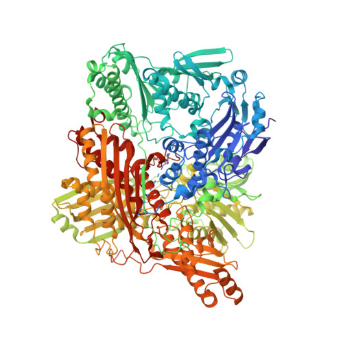
    Mammalian xanthine oxidoreductase can exist in both dehydrogenase and oxidase forms. Conversion between the two is implicated in such diverse processes as lactation, anti-bacterial activity, reperfusion injury and a growing number of diseases. We have constructed a variant of the rat liver enzyme that lacks the carboxy-terminal amino acids 1316-1331; it appears to assume an intermediate form, exhibiting a mixture of dehydrogenase and oxidase activities. The purified variant protein retained ~ 50-70% of oxidase activity even after prolonged dithiothreitol treatment, supporting a previous prediction that the C-terminal region plays a role in the dehydrogenase to oxidase conversion. In the crystal structure of the protein variant, most of the enzyme stays in an oxidase conformation. After 15 min of incubation with a high concentration of NADH, however, the corresponding X-ray structures showed a dehydrogenase-type conformation. On the other hand, disulfide formation between Cys535 and Cys992, which can clearly be seen in the electron density map of the crystal structure of the variant after removal of dithiothreitol, goes in parallel with the complete conversion to oxidase, resulting in structural changes identical to those observed upon proteolytic cleavage of the linker peptide. These results indicate that the dehydrogenase-oxidase transformation occurs rather readily and the insertion of the C-terminal peptide into the active site cavity of its subunit stabilizes the dehydrogenase form. We propose that the intermediate form can be generated (e.g. in endothelial cells) upon interaction of the C-terminal peptide portion of the enzyme with other proteins or the cell membrane.


  • Organizational Affiliation

    Department of Biochemistry and Molecular Biology, Nippon Medical School, Tokyo, Japan.


Macromolecules
Find similar proteins by:  (by identity cutoff)  |  3D Structure
Entity ID: 1
MoleculeChains Sequence LengthOrganismDetailsImage
Xanthine dehydrogenase/oxidase
A, B
1,315Rattus norvegicusMutation(s): 0 
Gene Names: Xdh
EC: 1.17.1.4 (PDB Primary Data), 1.17.3.2 (PDB Primary Data)
UniProt
Find proteins for P22985 (Rattus norvegicus)
Explore P22985 
Go to UniProtKB:  P22985
Entity Groups  
Sequence Clusters30% Identity50% Identity70% Identity90% Identity95% Identity100% Identity
UniProt GroupP22985
Sequence Annotations
Expand
  • Reference Sequence
Small Molecules
Ligands 6 Unique
IDChains Name / Formula / InChI Key2D Diagram3D Interactions
FAD
Query on FAD

Download Ideal Coordinates CCD File 
G [auth A],
M [auth B]
FLAVIN-ADENINE DINUCLEOTIDE
C27 H33 N9 O15 P2
VWWQXMAJTJZDQX-UYBVJOGSSA-N
FES
Query on FES

Download Ideal Coordinates CCD File 
C [auth A],
D [auth A],
J [auth B],
K [auth B]
FE2/S2 (INORGANIC) CLUSTER
Fe2 S2
NIXDOXVAJZFRNF-UHFFFAOYSA-N
URC
Query on URC

Download Ideal Coordinates CCD File 
H [auth A],
O [auth B]
URIC ACID
C5 H4 N4 O3
LEHOTFFKMJEONL-UHFFFAOYSA-N
GOL
Query on GOL

Download Ideal Coordinates CCD File 
I [auth A],
P [auth B],
Q [auth B]
GLYCEROL
C3 H8 O3
PEDCQBHIVMGVHV-UHFFFAOYSA-N
BCT
Query on BCT

Download Ideal Coordinates CCD File 
E [auth A],
N [auth B]
BICARBONATE ION
C H O3
BVKZGUZCCUSVTD-UHFFFAOYSA-M
CA
Query on CA

Download Ideal Coordinates CCD File 
F [auth A],
L [auth B]
CALCIUM ION
Ca
BHPQYMZQTOCNFJ-UHFFFAOYSA-N
Experimental Data & Validation

Experimental Data

  • Method: X-RAY DIFFRACTION
  • Resolution: 1.99 Å
  • R-Value Free: 0.229 
  • R-Value Work: 0.186 
  • R-Value Observed: 0.188 
  • Space Group: P 21 21 21
Unit Cell:
Length ( Å )Angle ( ˚ )
a = 98.497α = 90
b = 138.468β = 90
c = 222.213γ = 90
Software Package:
Software NamePurpose
REFMACrefinement
SCALEPACKdata scaling
PDB_EXTRACTdata extraction
SCALEPACKdata reduction
MOLREPphasing

Structure Validation

View Full Validation Report


Ligand Structure Quality Assessment 


Entry History 

Deposition Data

  • Released Date: 2015-04-29 
  • Deposition Author(s): Okamoto, K.

Revision History  (Full details and data files)

  • Version 1.0: 2015-04-29
    Type: Initial release
  • Version 1.1: 2015-09-16
    Changes: Database references
  • Version 1.2: 2020-02-05
    Changes: Data collection, Derived calculations
  • Version 1.3: 2023-11-08
    Changes: Data collection, Database references, Refinement description