4YNT

Crystal structure of Aspergillus flavus FAD glucose dehydrogenase


Experimental Data Snapshot

  • Method: X-RAY DIFFRACTION
  • Resolution: 1.78 Å
  • R-Value Free: 0.199 
  • R-Value Work: 0.153 
  • R-Value Observed: 0.156 

wwPDB Validation   3D Report Full Report


Ligand Structure Quality Assessment 


This is version 1.3 of the entry. See complete history


Literature

Structural analysis of fungus-derived FAD glucose dehydrogenase

Yoshida, H.Sakai, G.Mori, K.Kojima, K.Kamitori, S.Sode, K.

(2015) Sci Rep 5: 13498-13498

  • DOI: https://doi.org/10.1038/srep13498
  • Primary Citation of Related Structures:  
    4YNT, 4YNU

  • PubMed Abstract: 

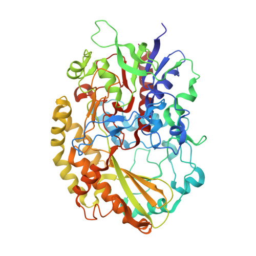
    We report the first three-dimensional structure of fungus-derived glucose dehydrogenase using flavin adenine dinucleotide (FAD) as the cofactor. This is currently the most advanced and popular enzyme used in glucose sensor strips manufactured for glycemic control by diabetic patients. We prepared recombinant nonglycosylated FAD-dependent glucose dehydrogenase (FADGDH) derived from Aspergillus flavus (AfGDH) and obtained the X-ray structures of the binary complex of enzyme and reduced FAD at a resolution of 1.78 Å and the ternary complex with reduced FAD and D-glucono-1,5-lactone (LGC) at a resolution of 1.57 Å. The overall structure is similar to that of fungal glucose oxidases (GOxs) reported till date. The ternary complex with reduced FAD and LGC revealed the residues recognizing the substrate. His505 and His548 were subjected for site-directed mutagenesis studies, and these two residues were revealed to form the catalytic pair, as those conserved in GOxs. The absence of residues that recognize the sixth hydroxyl group of the glucose of AfGDH, and the presence of significant cavity around the active site may account for this enzyme activity toward xylose. The structural information will contribute to the further engineering of FADGDH for use in more reliable and economical biosensing technology for diabetes management.


  • Organizational Affiliation

    Life Science Research Center and Faculty of Medicine, 1750-1, Ikenobe, Miki-cho, Kita-gun, Kagawa University, Kagawa 761-0793, Japan.


Macromolecules
Find similar proteins by:  (by identity cutoff)  |  3D Structure
Entity ID: 1
MoleculeChains Sequence LengthOrganismDetailsImage
Glucose oxidase, putative571Aspergillus flavus NRRL3357Mutation(s): 0 
Gene Names: AFLA_076820
EC: 1.1.5.9
UniProt
Find proteins for B8MX95 (Aspergillus flavus (strain ATCC 200026 / FGSC A1120 / IAM 13836 / NRRL 3357 / JCM 12722 / SRRC 167))
Explore B8MX95 
Go to UniProtKB:  B8MX95
Entity Groups  
Sequence Clusters30% Identity50% Identity70% Identity90% Identity95% Identity100% Identity
UniProt GroupB8MX95
Sequence Annotations
Expand
  • Reference Sequence
Small Molecules
Ligands 1 Unique
IDChains Name / Formula / InChI Key2D Diagram3D Interactions
FDA
Query on FDA

Download Ideal Coordinates CCD File 
B [auth A]DIHYDROFLAVINE-ADENINE DINUCLEOTIDE
C27 H35 N9 O15 P2
YPZRHBJKEMOYQH-UYBVJOGSSA-N
Experimental Data & Validation

Experimental Data

  • Method: X-RAY DIFFRACTION
  • Resolution: 1.78 Å
  • R-Value Free: 0.199 
  • R-Value Work: 0.153 
  • R-Value Observed: 0.156 
  • Space Group: P 1 21 1
Unit Cell:
Length ( Å )Angle ( ˚ )
a = 49.907α = 90
b = 64.359β = 101.43
c = 76.166γ = 90
Software Package:
Software NamePurpose
REFMACrefinement
SCALEPACKdata reduction
PDB_EXTRACTdata extraction
HKL-2000data scaling
PHENIXphasing

Structure Validation

View Full Validation Report


Ligand Structure Quality Assessment 


Entry History 

Deposition Data

Revision History  (Full details and data files)

  • Version 1.0: 2015-09-02
    Type: Initial release
  • Version 1.1: 2015-09-16
    Changes: Database references
  • Version 1.2: 2020-02-05
    Changes: Data collection, Derived calculations
  • Version 1.3: 2023-11-08
    Changes: Data collection, Database references, Refinement description