4YIG

vaccinia virus D4/A20(1-50) in complex with dsDNA containing an abasic site and free uracyl


Experimental Data Snapshot

  • Method: X-RAY DIFFRACTION
  • Resolution: 2.70 Å
  • R-Value Free: 0.228 
  • R-Value Work: 0.180 
  • R-Value Observed: 0.182 

wwPDB Validation   3D Report Full Report


This is version 2.1 of the entry. See complete history


Literature

Crystal Structure of the Vaccinia Virus Uracil-DNA Glycosylase in Complex with DNA.

Burmeister, W.P.Tarbouriech, N.Fender, P.Contesto-Richefeu, C.Peyrefitte, C.N.Iseni, F.

(2015) J Biol Chem 290: 17923-17934

  • DOI: https://doi.org/10.1074/jbc.M115.648352
  • Primary Citation of Related Structures:  
    4YGM, 4YIG

  • PubMed Abstract: 

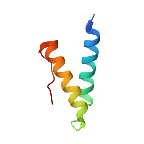
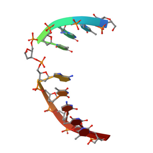
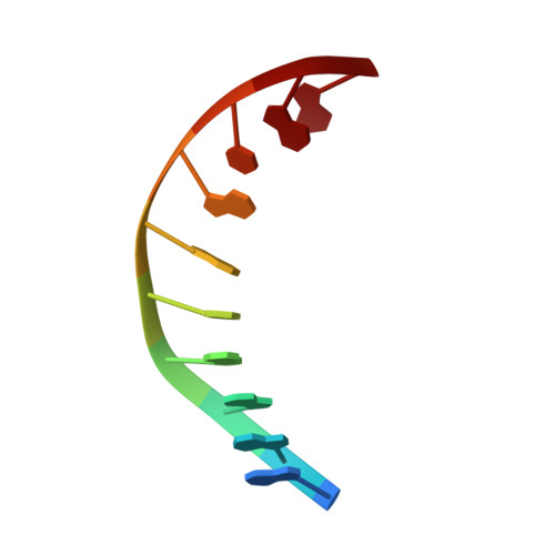
    Vaccinia virus polymerase holoenzyme is composed of the DNA polymerase catalytic subunit E9 associated with its heterodimeric co-factor A20·D4 required for processive genome synthesis. Although A20 has no known enzymatic activity, D4 is an active uracil-DNA glycosylase (UNG). The presence of a repair enzyme as a component of the viral replication machinery suggests that, for poxviruses, DNA synthesis and base excision repair is coupled. We present the 2.7 Å crystal structure of the complex formed by D4 and the first 50 amino acids of A20 (D4·A201-50) bound to a 10-mer DNA duplex containing an abasic site resulting from the cleavage of a uracil base. Comparison of the viral complex with its human counterpart revealed major divergences in the contacts between protein and DNA and in the enzyme orientation on the DNA. However, the conformation of the dsDNA within both structures is very similar, suggesting a dominant role of the DNA conformation for UNG function. In contrast to human UNG, D4 appears rigid, and we do not observe a conformational change upon DNA binding. We also studied the interaction of D4·A201-50 with different DNA oligomers by surface plasmon resonance. D4 binds weakly to nonspecific DNA and to uracil-containing substrates but binds abasic sites with a Kd of <1.4 μm. This second DNA complex structure of a family I UNG gives new insight into the role of D4 as a co-factor of vaccinia virus DNA polymerase and allows a better understanding of the structural determinants required for UNG action.


  • Organizational Affiliation

    Université Grenoble Alpes, Unit of Virus Host Cell Interactions (UVHCI), F-38000 Grenoble, France; CNRS, UVHCI, F-38000 Grenoble, France. Electronic address: wim.burmeister@ujf-grenoble.fr.


Macromolecules

Find similar proteins by:  (by identity cutoff)  |  3D Structure
Entity ID: 1
MoleculeChains Sequence LengthOrganismDetailsImage
Uracil-DNA glycosylase
A, E, I
232Vaccinia virus CopenhagenMutation(s): 0 
Gene Names: UNGD4R
EC: 3.2.2.27
UniProt
Find proteins for P20536 (Vaccinia virus (strain Copenhagen))
Explore P20536 
Go to UniProtKB:  P20536
Entity Groups  
Sequence Clusters30% Identity50% Identity70% Identity90% Identity95% Identity100% Identity
UniProt GroupP20536
Sequence Annotations
Expand
  • Reference Sequence
Find similar proteins by:  (by identity cutoff)  |  3D Structure
Entity ID: 2
MoleculeChains Sequence LengthOrganismDetailsImage
DNA polymerase processivity factor component A20
B, F, J
52Vaccinia virus CopenhagenMutation(s): 0 
Gene Names: A20R
UniProt
Find proteins for P20995 (Vaccinia virus (strain Copenhagen))
Explore P20995 
Go to UniProtKB:  P20995
Entity Groups  
Sequence Clusters30% Identity50% Identity70% Identity90% Identity95% Identity100% Identity
UniProt GroupP20995
Sequence Annotations
Expand
  • Reference Sequence

Find similar nucleic acids by:  Sequence   |   3D Structure  

Entity ID: 3
MoleculeChains LengthOrganismImage
DNA (5'-D(*CP*TP*GP*TP*(ORP)P*AP*TP*CP*TP*T)-3')
C, G, K
10synthetic construct
Sequence Annotations
Expand
  • Reference Sequence

Find similar nucleic acids by:  Sequence   |   3D Structure  

Entity ID: 4
MoleculeChains LengthOrganismImage
DNA (5'-D(*AP*AP*GP*AP*TP*AP*AP*CP*AP*G)-3')
D, H, L
10synthetic construct
Sequence Annotations
Expand
  • Reference Sequence
Experimental Data & Validation

Experimental Data

  • Method: X-RAY DIFFRACTION
  • Resolution: 2.70 Å
  • R-Value Free: 0.228 
  • R-Value Work: 0.180 
  • R-Value Observed: 0.182 
  • Space Group: P 65
Unit Cell:
Length ( Å )Angle ( ˚ )
a = 136.092α = 90
b = 136.092β = 90
c = 161.073γ = 120
Software Package:
Software NamePurpose
REFMACrefinement
XDSdata reduction
Aimlessdata scaling
PHASERphasing

Structure Validation

View Full Validation Report


Entry History & Funding Information

Deposition Data


Funding OrganizationLocationGrant Number
French National Research AgencyFranceanr-13-bsv8-0014

Revision History  (Full details and data files)

  • Version 1.0: 2015-06-10
    Type: Initial release
  • Version 1.1: 2015-06-17
    Changes: Database references
  • Version 1.2: 2015-07-29
    Changes: Database references
  • Version 2.0: 2017-09-06
    Changes: Atomic model, Author supporting evidence, Derived calculations
  • Version 2.1: 2024-01-10
    Changes: Data collection, Database references, Refinement description