


Pilz domain with c-di-gmp of a protein from Pseudomonas aeruginosa
Chi, K.K., Yuan, Z.L.To be published.
Experimental Data Snapshot
Entity ID: 1 | |||||
|---|---|---|---|---|---|
| Molecule | Chains | Sequence Length | Organism | Details | Image |
| Alginate biosynthesis protein Alg44 | 111 | Pseudomonas aeruginosa PAO1 | Mutation(s): 0 Gene Names: alg44, PA3542 | 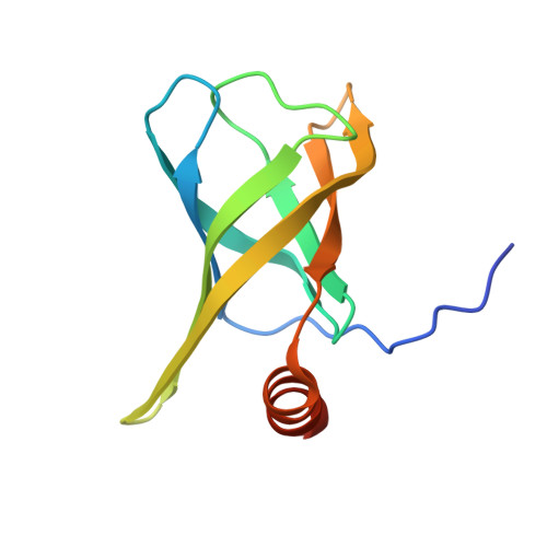 | |
UniProt | |||||
Find proteins for Q9HY69 (Pseudomonas aeruginosa (strain ATCC 15692 / DSM 22644 / CIP 104116 / JCM 14847 / LMG 12228 / 1C / PRS 101 / PAO1)) Explore Q9HY69 Go to UniProtKB: Q9HY69 | |||||
Entity Groups | |||||
| Sequence Clusters | 30% Identity50% Identity70% Identity90% Identity95% Identity100% Identity | ||||
| UniProt Group | Q9HY69 | ||||
Sequence AnnotationsExpand | |||||
| |||||
| Ligands 2 Unique | |||||
|---|---|---|---|---|---|
| ID | Chains | Name / Formula / InChI Key | 2D Diagram | 3D Interactions | |
| C2E Query on C2E | E [auth A] F [auth A] G [auth A] J [auth B] K [auth B] | 9,9'-[(2R,3R,3aS,5S,7aR,9R,10R,10aS,12S,14aR)-3,5,10,12-tetrahydroxy-5,12-dioxidooctahydro-2H,7H-difuro[3,2-d:3',2'-j][1,3,7,9,2,8]tetraoxadiphosphacyclododecine-2,9-diyl]bis(2-amino-1,9-dihydro-6H-purin-6-one) C20 H24 N10 O14 P2 PKFDLKSEZWEFGL-MHARETSRSA-N |  | ||
| ZN Query on ZN | H [auth A] I [auth A] M [auth B] N [auth B] O [auth B] | ZINC ION Zn PTFCDOFLOPIGGS-UHFFFAOYSA-N |  | ||
| Length ( Å ) | Angle ( ˚ ) |
|---|---|
| a = 75.526 | α = 90 |
| b = 75.526 | β = 90 |
| c = 195.37 | γ = 90 |
| Software Name | Purpose |
|---|---|
| PHENIX | refinement |
| HKL-2000 | data reduction |
| HKL-2000 | data scaling |
| PHENIX | phasing |
RCSB PDB (citation) is hosted by
RCSB PDB is a member of the

