4XJ6

Crystal structure of Escherichia coli DncV 3'-deoxy GTP bound form


Experimental Data Snapshot

  • Method: X-RAY DIFFRACTION
  • Resolution: 2.31 Å
  • R-Value Free: 0.234 
  • R-Value Work: 0.189 
  • R-Value Observed: 0.191 

wwPDB Validation   3D Report Full Report


Ligand Structure Quality Assessment 


This is version 1.3 of the entry. See complete history


Literature

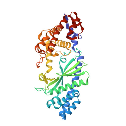
Structural Basis for the Catalytic Mechanism of DncV, Bacterial Homolog of Cyclic GMP-AMP Synthase

Kato, K.Ishii, R.Hirano, S.Ishitani, R.Nureki, O.

(2015) Structure 23: 843-850

  • DOI: https://doi.org/10.1016/j.str.2015.01.023
  • Primary Citation of Related Structures:  
    4XJ1, 4XJ3, 4XJ4, 4XJ5, 4XJ6

  • PubMed Abstract: 

    Cyclic dinucleotides (CDNs) play key roles as second messengers and signaling molecules in bacteria and metazoans. The newly identified dinucleotide cyclase in Vibrio cholerae (DncV) produces three different CDNs containing two 3'-5' phosphodiester bonds, and its predominant product is cyclic GMP-AMP, whereas mammalian cyclic GMP-AMP synthase (cGAS) produces only cyclic GMP-AMP containing mixed 2'-5' phosphodiester bonds. We report the crystal structures of V. cholerae and Escherichia coli DncV in complex with various nucleotides in the pre-reaction states. The high-resolution structures revealed that DncV preferably recognizes ATP and GTP as acceptor and donor nucleotides, respectively, in the first nucleotidyl transfer reaction. Considering the recently reported intermediate structures, our pre-reaction state structures provide the precise mechanism of 3'-5' linked cyclic AMP-GMP production in bacteria. A comparison with cGAS in the pre-reaction states suggests that the orientation of the acceptor nucleotide primarily determines the distinct linkage specificities between DncV and cGAS.


  • Organizational Affiliation

    Department of Biological Sciences, Graduate School of Science, The University of Tokyo, Bunkyo-ku, Tokyo 113-0032, Japan.


Macromolecules
Find similar proteins by:  (by identity cutoff)  |  3D Structure
Entity ID: 1
MoleculeChains Sequence LengthOrganismDetailsImage
VC0179-like protein407Escherichia coliMutation(s): 0 
UniProt
Find proteins for Q6XGD8 (Escherichia coli)
Explore Q6XGD8 
Go to UniProtKB:  Q6XGD8
Entity Groups  
Sequence Clusters30% Identity50% Identity70% Identity90% Identity95% Identity100% Identity
UniProt GroupQ6XGD8
Sequence Annotations
Expand
  • Reference Sequence
Experimental Data & Validation

Experimental Data

  • Method: X-RAY DIFFRACTION
  • Resolution: 2.31 Å
  • R-Value Free: 0.234 
  • R-Value Work: 0.189 
  • R-Value Observed: 0.191 
  • Space Group: C 1 2 1
Unit Cell:
Length ( Å )Angle ( ˚ )
a = 136.07α = 90
b = 46.3β = 110.61
c = 62.56γ = 90
Software Package:
Software NamePurpose
PHENIXrefinement
XDSdata reduction
MOLREPphasing

Structure Validation

View Full Validation Report


Ligand Structure Quality Assessment 


Entry History 

Deposition Data

Revision History  (Full details and data files)

  • Version 1.0: 2015-04-29
    Type: Initial release
  • Version 1.1: 2015-05-20
    Changes: Database references
  • Version 1.2: 2020-02-05
    Changes: Data collection, Database references, Derived calculations, Source and taxonomy, Structure summary
  • Version 1.3: 2024-03-20
    Changes: Data collection, Database references, Derived calculations