4XHC

rhamnosidase from Klebsiella oxytoca with rhamnose bound


Experimental Data Snapshot

  • Method: X-RAY DIFFRACTION
  • Resolution: 2.70 Å
  • R-Value Free: 0.202 
  • R-Value Work: 0.182 
  • R-Value Observed: 0.183 

wwPDB Validation   3D Report Full Report


Ligand Structure Quality Assessment 


This is version 2.1 of the entry. See complete history


Literature

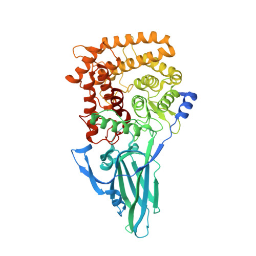
Crystal structure of a novel two domain GH78 family alpha-rhamnosidase from Klebsiella oxytoca with rhamnose bound.

O'Neill, E.C.Stevenson, C.E.Paterson, M.J.Rejzek, M.Chauvin, A.L.Lawson, D.M.Field, R.A.

(2015) Proteins 83: 1742-1749

  • DOI: https://doi.org/10.1002/prot.24807
  • Primary Citation of Related Structures:  
    4XHC

  • PubMed Abstract: 

    The crystal structure of the GH78 family α-rhamnosidase from Klebsiella oxytoca (KoRha) has been determined at 2.7 Å resolution with rhamnose bound in the active site of the catalytic domain. Curiously, the putative catalytic acid, Asp 222, is preceded by an unusual non-proline cis-peptide bond which helps to project the carboxyl group into the active centre. This KoRha homodimeric structure is significantly smaller than those of the other previously determined GH78 structures. Nevertheless, the enzyme displays α-rhamnosidase activity when assayed in vitro, suggesting that the additional structural domains found in the related enzymes are dispensible for function.


  • Organizational Affiliation

    Department of Biological Chemistry, John Innes Centre, Norwich Research Park, Norwich, Nr4 7UH, United Kingdom.


Macromolecules
Find similar proteins by:  (by identity cutoff)  |  3D Structure
Entity ID: 1
MoleculeChains Sequence LengthOrganismDetailsImage
Alpha-L-rhamnosidase
A, B
543Klebsiella oxytocaMutation(s): 0 
Gene Names: KOX_19945
EC: 3.2.1.40
UniProt
Find proteins for A0A0J9X262 (Klebsiella oxytoca)
Explore A0A0J9X262 
Go to UniProtKB:  A0A0J9X262
Entity Groups  
Sequence Clusters30% Identity50% Identity70% Identity90% Identity95% Identity100% Identity
UniProt GroupA0A0J9X262
Sequence Annotations
Expand
  • Reference Sequence
Experimental Data & Validation

Experimental Data

  • Method: X-RAY DIFFRACTION
  • Resolution: 2.70 Å
  • R-Value Free: 0.202 
  • R-Value Work: 0.182 
  • R-Value Observed: 0.183 
  • Space Group: P 41 21 2
Unit Cell:
Length ( Å )Angle ( ˚ )
a = 148.43α = 90
b = 148.43β = 90
c = 202.16γ = 90
Software Package:
Software NamePurpose
SHELXphasing
REFMACrefinement
PDB_EXTRACTdata extraction
Aimlessdata scaling

Structure Validation

View Full Validation Report


Ligand Structure Quality Assessment 


Entry History & Funding Information

Deposition Data


Funding OrganizationLocationGrant Number
Biotechnology and Biological Sciences Research CouncilUnited KingdomBB/J004561/1

Revision History  (Full details and data files)

  • Version 1.0: 2015-04-15
    Type: Initial release
  • Version 1.1: 2015-04-22
    Changes: Database references
  • Version 1.2: 2015-09-02
    Changes: Database references
  • Version 2.0: 2017-08-30
    Changes: Atomic model, Author supporting evidence
  • Version 2.1: 2020-07-29
    Type: Remediation
    Reason: Carbohydrate remediation
    Changes: Data collection, Derived calculations, Structure summary