Structural insights into the substrate specificity of two esterases from the thermophilic Rhizomucor miehei
Yang, S., Qin, Z., Duan, X., Yan, Q., Jiang, Z.(2015) J Lipid Res 56: 1616-1624
- PubMed: 26108223
- DOI: https://doi.org/10.1194/jlr.M060673
- Primary Citation of Related Structures:
4WY5, 4WY8 - PubMed Abstract:
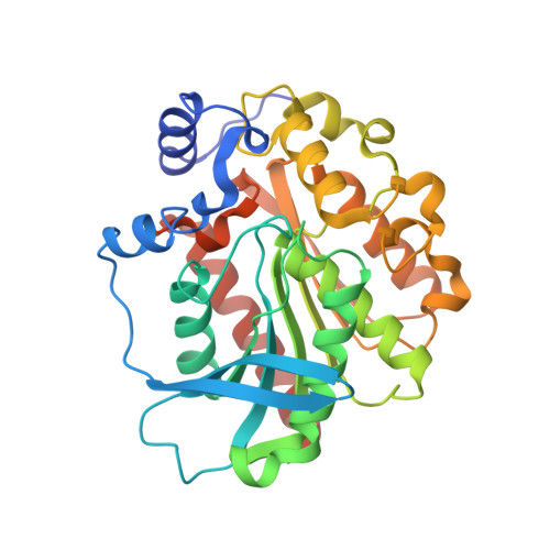
Two hormone-sensitive lipase (HSL) family esterases (RmEstA and RmEstB) from the thermophilic fungus Rhizomucor miehei, exhibiting distinct substrate specificity, have been recently reported to show great potential in industrial applications. In this study, the crystal structures of RmEstA and RmEstB were determined at 2.15 Å and 2.43 Å resolutions, respectively. The structures of RmEstA and RmEstB showed two distinctive domains, a catalytic domain and a cap domain, with the classical α/β-hydrolase fold. Catalytic triads consisting of residues Ser161, Asp262, and His292 in RmEstA, and Ser164, Asp261, and His291 in RmEstB were found in the respective canonical positions. Structural comparison of RmEstA and RmEstB revealed that their distinct substrate specificity might be attributed to their different substrate-binding pockets. The aromatic amino acids Phe222 and Trp92, located in the center of the substrate-binding pocket of RmEstB, blocked this pocket, thus narrowing its catalytic range for substrates (C2-C8). Two mutants (F222A and W92F in RmEstB) showing higher catalytic activity toward long-chain substrates further confirmed the hypothesized interference. This is the first report of HSL family esterase structures from filamentous fungi. The information on structure-function relationships could open important avenues of exploration for further industrial applications of esterases.
Organizational Affiliation:
College of Food Science and Nutritional Engineering, The Research and Innovation Center of Food Nutrition and Human Health (Beijing), China Agricultural University, Beijing 100083, China.