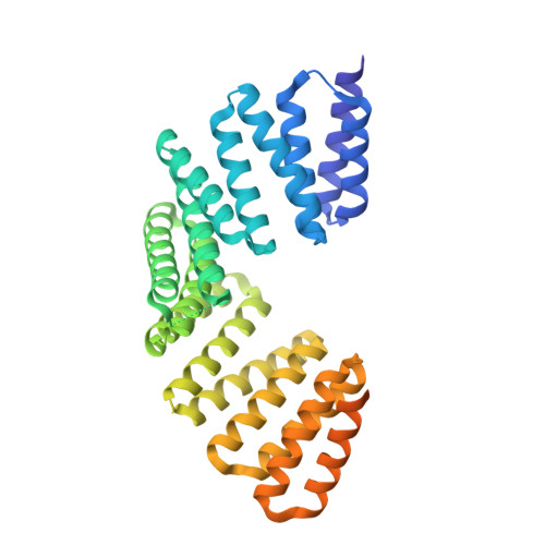
4WNE

Crystal structure of the TPR domain of LGN in complex with Frmpd4/Preso1 at 2.0 Angstrom resolution


Experimental Data Snapshot

  • Method: X-RAY DIFFRACTION
  • Resolution: 2.00 Å
  • R-Value Free: 0.238 
  • R-Value Work: 0.210 
  • R-Value Observed: 0.212 

wwPDB Validation   3D Report Full Report


This is version 1.2 of the entry. See complete history


Literature

Structural basis for the recognition of the scaffold protein Frmpd4/Preso1 by the TPR domain of the adaptor protein LGN

Takayanagi, H.Yuzawa, S.Sumimoto, H.

(2015) Acta Crystallogr F Struct Biol Commun 71: 175-183

  • DOI: https://doi.org/10.1107/S2053230X14028143
  • Primary Citation of Related Structures:  
    4WND, 4WNE, 4WNF, 4WNG

  • PubMed Abstract: 

    The adaptor protein LGN interacts via the N-terminal domain comprising eight tetratricopeptide-repeat (TPR) motifs with its partner proteins mInsc, NuMA, Frmpd1 and Frmpd4 in a mutually exclusive manner. Here, the crystal structure of the LGN TPR domain in complex with human Frmpd4 is described at 1.5 Å resolution. In the complex, the LGN-binding region of Frmpd4 (amino-acid residues 990-1011) adopts an extended structure that runs antiparallel to LGN along the concave surface of the superhelix formed by the TPR motifs. Comparison with the previously determined structures of the LGN-Frmpd1, LGN-mInsc and LGN-NuMA complexes reveals that these partner proteins interact with LGN TPR1-6 via a common core binding region with consensus sequence (E/Q)XEX4-5(E/D/Q)X1-2(K/R)X0-1(V/I). In contrast to Frmpd1, Frmpd4 makes additional contacts with LGN via regions N- and C-terminal to the core sequence. The N-terminal extension is replaced by a specific α-helix in mInsc, which drastically increases the direct contacts with LGN TPR7/8, consistent with the higher affinity of mInsc for LGN. A crystal structure of Frmpd4-bound LGN in an oxidized form is also reported, although oxidation does not appear to strongly affect the interaction with Frmpd4.


  • Organizational Affiliation

    Department of Biochemistry, Kyushu University Graduate School of Medical Sciences, 3-1-1 Maidashi, Higashi-ku, Fukuoka 812-8582, Japan.


Macromolecules
Find similar proteins by:  (by identity cutoff)  |  3D Structure
Entity ID: 1
MoleculeChains Sequence LengthOrganismDetailsImage
G-protein-signaling modulator 2406Homo sapiensMutation(s): 0 
Gene Names: GPSM2LGN
UniProt & NIH Common Fund Data Resources
Find proteins for P81274 (Homo sapiens)
Explore P81274 
Go to UniProtKB:  P81274
PHAROS:  P81274
GTEx:  ENSG00000121957 
Entity Groups  
Sequence Clusters30% Identity50% Identity70% Identity90% Identity95% Identity100% Identity
UniProt GroupP81274
Sequence Annotations
Expand
  • Reference Sequence

Find similar proteins by:  Sequence   |   3D Structure  

Entity ID: 2
MoleculeChains Sequence LengthOrganismDetailsImage
Peptide from FERM and PDZ domain-containing protein 425Homo sapiensMutation(s): 0 
UniProt & NIH Common Fund Data Resources
Find proteins for Q14CM0 (Homo sapiens)
Explore Q14CM0 
Go to UniProtKB:  Q14CM0
PHAROS:  Q14CM0
GTEx:  ENSG00000169933 
Entity Groups  
Sequence Clusters30% Identity50% Identity70% Identity90% Identity95% Identity100% Identity
UniProt GroupQ14CM0
Sequence Annotations
Expand
  • Reference Sequence
Experimental Data & Validation

Experimental Data

  • Method: X-RAY DIFFRACTION
  • Resolution: 2.00 Å
  • R-Value Free: 0.238 
  • R-Value Work: 0.210 
  • R-Value Observed: 0.212 
  • Space Group: P 61 2 2
Unit Cell:
Length ( Å )Angle ( ˚ )
a = 91.784α = 90
b = 91.784β = 90
c = 176.783γ = 120
Software Package:
Software NamePurpose
HKL-2000data reduction
HKL-2000data scaling
MOLREPphasing
Cootmodel building
REFMACrefinement
PDB_EXTRACTdata extraction

Structure Validation

View Full Validation Report


Entry History 

Deposition Data

Revision History  (Full details and data files)

  • Version 1.0: 2015-09-16
    Type: Initial release
  • Version 1.1: 2020-02-05
    Changes: Data collection, Derived calculations, Refinement description
  • Version 1.2: 2023-11-08
    Changes: Data collection, Database references, Refinement description