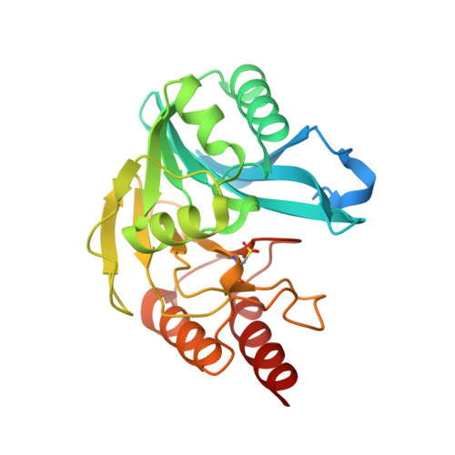
Structural and Biochemical Characterization of Vim-26 Show that Leu224 Has Implications for the Substrate Specificity of Vim Metallo-Beta-Lactamases.
Leiros, H.S., Edvardsen, K.S.W., Bjerga, G.E.K., Samuelsen, O.(2015) FEBS J 282: 1031
- PubMed: 25601024
- DOI: https://doi.org/10.1111/febs.13200
- Primary Citation of Related Structures:
4UWO, 4UWP, 4UWR, 4UWS - PubMed Abstract:
During the last decades antimicrobial resistance has become a global health problem. Metallo-β-lactamases (MBLs) which are broad-spectrum β-lactamases that inactivate virtually all β-lactams including carbapenems, are contributing to this health problem. In this study a novel MBL variant, termed VIM-26, identified in a Klebsiella pneumoniae isolate was studied. VIM-26 belongs to the Verona integron-encoded metallo-β-lactamase (VIM) family of MBLs and is a His224Leu variant of the well-characterized VIM-1 variant. In this study, we report the kinetic parameters, minimum inhibitory concentrations and crystal structures of a recombinant VIM-26 protein, and compare them to previously published data on VIM-1, VIM-2 and VIM-7. The kinetic parameters and minimum inhibitory concentration determinations show that VIM-26, like VIM-7, has higher penicillinase activity but lower cephalosporinase activity than VIM-1 and VIM-2. The four determined VIM-26 crystal structures revealed mono- and di-zinc forms, where the Zn1 ion has distorted tetrahedral coordination geometry with an additional water molecule (W2) at a distance of 2.6-3.7 Å, which could be important during catalysis. The R2 drug binding site in VIM-26 is more open compared to VIM-2 and VIM-7 and neutrally charged due to Leu224 and Ser228. Thus, the VIM-26 drug binding properties are different from the VIM-2 (Tyr224/Arg228) and VIM-7 (His224/Arg228) structures, indicating a role of these residues in the substrate specificity.
Organizational Affiliation:
Norwegian Structural Biology Centre, Department of Chemistry, UiT The Arctic University of Norway, Tromsø, Norway.