Structural Flexibility of the Heme Cavity in the Cold-Adapted Truncated Hemoglobin from the Antarctic Marine Bacterium Pseudoalteromonas Haloplanktis Tac125.
Giordano, D., Pesce, A., Boechi, L., Bustamante, J.P., Caldelli, E., Howes, B.D., Riccio, A., Di Prisco, G., Nardini, M., Estrin, D., Smulevich, G., Bolognesi, M., Verde, C.(2015) FEBS J 282: 2948
- PubMed: 26040838
- DOI: https://doi.org/10.1111/febs.13335
- Primary Citation of Related Structures:
4UUR - PubMed Abstract:
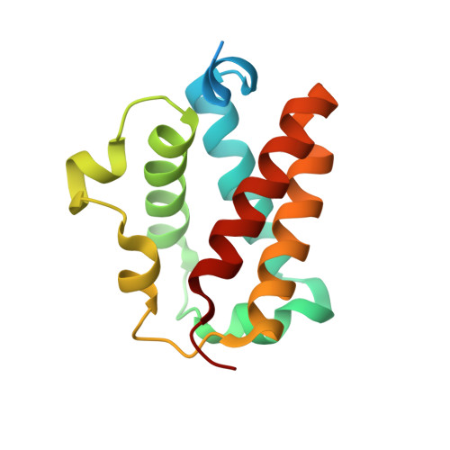
Truncated hemoglobins build one of the three branches of the globin protein superfamily. They display a characteristic two-on-two α-helical sandwich fold and are clustered into three groups (I, II and III) based on distinct structural features. Truncated hemoglobins are present in eubacteria, cyanobacteria, protozoa and plants. Here we present a structural, spectroscopic and molecular dynamics characterization of a group-II truncated hemoglobin, encoded by the PSHAa0030 gene from Pseudoalteromonas haloplanktis TAC125 (Ph-2/2HbO), a cold-adapted Antarctic marine bacterium hosting one flavohemoglobin and three distinct truncated hemoglobins. The Ph-2/2HbO aquo-met crystal structure (at 2.21 Å resolution) shows typical features of group-II truncated hemoglobins, namely the two-on-two α-helical sandwich fold, a helix Φ preceding the proximal helix F, and a heme distal-site hydrogen-bonded network that includes water molecules and several distal-site residues, including His(58)CD1. Analysis of Ph-2/2HbO by electron paramagnetic resonance, resonance Raman and electronic absorption spectra, under varied solution conditions, shows that Ph-2/2HbO can access diverse heme ligation states. Among these, detection of a low-spin heme hexa-coordinated species suggests that residue Tyr(42)B10 can undergo large conformational changes in order to act as the sixth heme-Fe ligand. Altogether, the results show that Ph-2/2HbO maintains the general structural features of group-II truncated hemoglobins but displays enhanced conformational flexibility in the proximity of the heme cavity, a property probably related to the functional challenges, such as low temperature, high O2 concentration and low kinetic energy of molecules, experienced by organisms living in the Antarctic environment.
Organizational Affiliation:
Institute of Biosciences and BioResources, National Research Council, Napoli, Italy.