4TL3

Mechanistic insights from the crystal structure of an inward proton-transporting Anabaena sensory rhodopsin mutant

  • Classification: SIGNALING PROTEIN
  • Organism(s): Nostoc sp. PCC 7120 = FACHB-418
  • Expression System: Escherichia coli
  • Mutation(s): Yes 
  • Membrane Protein: Yes  OPMPDBTMMemProtMD

  • Deposited: 2014-05-29 Released: 2014-07-16 
  • Deposition Author(s): Dong, B.H., Luecke, H.
  • Funding Organization(s): National Institutes of Health/National Institute of General Medical Sciences (NIH/NIGMS), National Institutes of Health/National Institute Of Allergy and Infectious Diseases (NIH/NIAID), UC Irvine Center for Biomembrane Systems

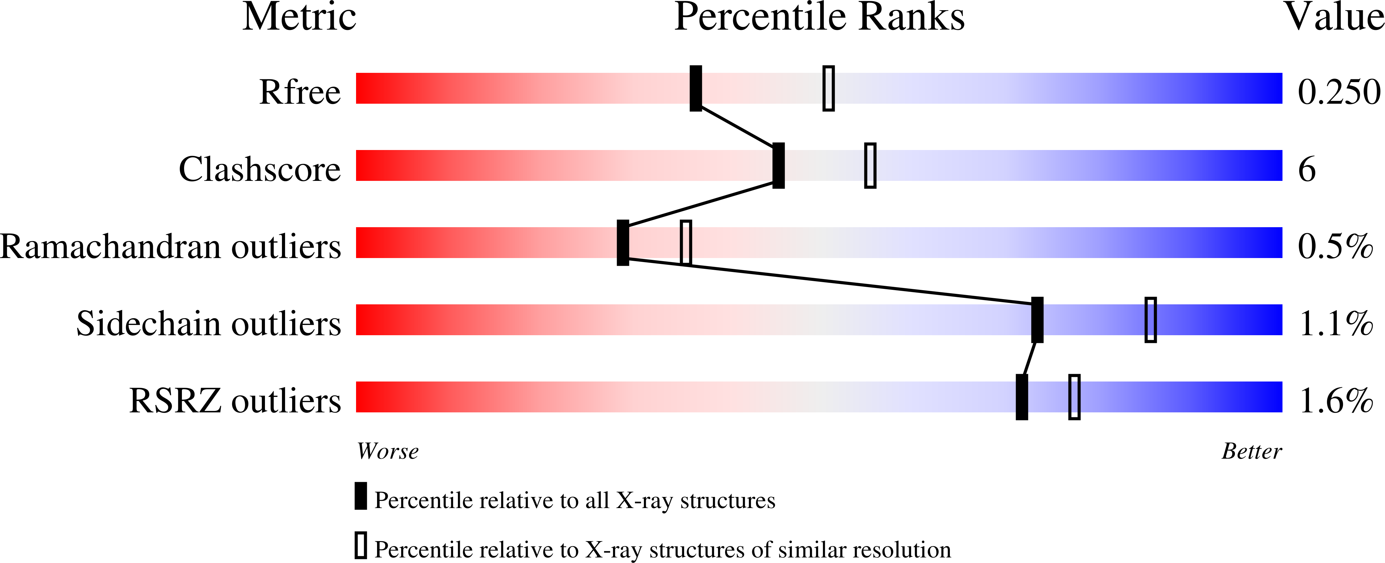
Experimental Data Snapshot

  • Method: X-RAY DIFFRACTION
  • Resolution: 2.30 Å
  • R-Value Free: 0.250 
  • R-Value Work: 0.220 
  • R-Value Observed: 0.221 

wwPDB Validation   3D Report Full Report


Ligand Structure Quality Assessment 


This is version 2.0 of the entry. See complete history


Literature

Mechanistic insights from the crystal structure of an inward proton-transportingAnabaena sensory rhodopsin mutant

Dong, B.H.Luecke, H.

To be published.

Macromolecules
Find similar proteins by:  (by identity cutoff)  |  3D Structure
Entity ID: 1
MoleculeChains Sequence LengthOrganismDetailsImage
Anabaena sensory rhodopsin
A, B
226Nostoc sp. PCC 7120 = FACHB-418Mutation(s): 1 
Gene Names: alr3165
Membrane Entity: Yes 
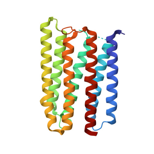
UniProt
Find proteins for Q8YSC4 (Nostoc sp. (strain PCC 7120 / SAG 25.82 / UTEX 2576))
Explore Q8YSC4 
Go to UniProtKB:  Q8YSC4
Entity Groups  
Sequence Clusters30% Identity50% Identity70% Identity90% Identity95% Identity100% Identity
UniProt GroupQ8YSC4
Sequence Annotations
Expand
  • Reference Sequence
Small Molecules
Ligands 2 Unique
IDChains Name / Formula / InChI Key2D Diagram3D Interactions
PEE
Query on PEE

Download Ideal Coordinates CCD File 
AA [auth B]
BA [auth B]
CA [auth B]
D [auth A]
DA [auth B]
AA [auth B],
BA [auth B],
CA [auth B],
D [auth A],
DA [auth B],
E [auth A],
EA [auth B],
F [auth A],
FA [auth B],
G [auth A],
GA [auth B],
H [auth A],
HA [auth B],
I [auth A],
IA [auth B],
J [auth A],
JA [auth B],
K [auth A],
KA [auth B],
L [auth A],
LA [auth B],
M [auth A],
MA [auth B],
N [auth A],
O [auth A],
P [auth A],
Q [auth A],
R [auth A],
S [auth A],
U [auth B],
V [auth B],
W [auth B],
X [auth B],
Y [auth B],
Z [auth B]
1,2-dioleoyl-sn-glycero-3-phosphoethanolamine
C41 H78 N O8 P
MWRBNPKJOOWZPW-NYVOMTAGSA-N
RET
Query on RET

Download Ideal Coordinates CCD File 
C [auth A],
T [auth B]
RETINAL
C20 H28 O
NCYCYZXNIZJOKI-OVSJKPMPSA-N
Experimental Data & Validation

Experimental Data

  • Method: X-RAY DIFFRACTION
  • Resolution: 2.30 Å
  • R-Value Free: 0.250 
  • R-Value Work: 0.220 
  • R-Value Observed: 0.221 
  • Space Group: P 21 21 21
Unit Cell:
Length ( Å )Angle ( ˚ )
a = 55.265α = 90
b = 104.301β = 90
c = 111.386γ = 90
Software Package:
Software NamePurpose
REFMACrefinement

Structure Validation

View Full Validation Report


Ligand Structure Quality Assessment 


Entry History & Funding Information

Deposition Data


Funding OrganizationLocationGrant Number
National Institutes of Health/National Institute of General Medical Sciences (NIH/NIGMS)United StatesR01 GM067808
National Institutes of Health/National Institute Of Allergy and Infectious Diseases (NIH/NIAID)United StatesAI078000
UC Irvine Center for Biomembrane SystemsUnited States--

Revision History  (Full details and data files)

  • Version 1.0: 2014-07-16
    Type: Initial release
  • Version 1.1: 2014-10-29
    Changes: Non-polymer description
  • Version 1.2: 2017-09-06
    Changes: Author supporting evidence, Derived calculations, Other, Source and taxonomy
  • Version 1.3: 2019-12-11
    Changes: Author supporting evidence
  • Version 2.0: 2023-09-27
    Changes: Advisory, Atomic model, Data collection, Database references, Derived calculations, Non-polymer description, Refinement description, Structure summary