4RVC

Structure of ATP binding subunit of ABC transporter


Experimental Data Snapshot

  • Method: X-RAY DIFFRACTION
  • Resolution: 1.77 Å
  • R-Value Free: 0.250 
  • R-Value Work: 0.216 
  • R-Value Observed: 0.250 

wwPDB Validation   3D Report Full Report


This is version 1.2 of the entry. See complete history


Literature

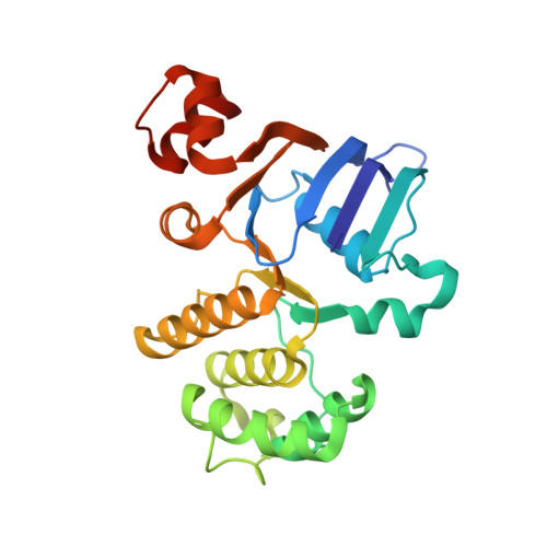
Crystal structure of ATP-binding subunit of an ABC transporter from Geobacillus kaustophilus.

Manjula, M.Pampa, K.J.Kumar, S.M.Mukherjee, S.Kunishima, N.Rangappa, K.S.Lokanath, N.K.

(2015) Biochem Biophys Res Commun 459: 113-117

  • DOI: https://doi.org/10.1016/j.bbrc.2015.02.079
  • Primary Citation of Related Structures:  
    4RVC

  • PubMed Abstract: 

    The ATP binding cassette (ABC) transporters, represent one of the largest superfamilies of primary transporters, which are very essential for various biological functions. The crystal structure of ATP-binding subunit of an ABC transporter from Geobacillus kaustophilus has been determined at 1.77 Å resolution. The crystal structure revealed that the protomer has two thick arms, (arm I and II), which resemble 'L' shape. The ATP-binding pocket is located close to the end of arm I. ATP molecule is docked into the active site of the protein. The dimeric crystal structure of ATP-binding subunit of ABC transporter from G. kaustophilus has been compared with the previously reported crystal structure of ATP-binding subunit of ABC transporter from Salmonella typhimurium.


  • Organizational Affiliation

    Department of Studies in Physics, University of Mysore, Mysore 570 006, India.


Macromolecules
Find similar proteins by:  (by identity cutoff)  |  3D Structure
Entity ID: 1
MoleculeChains Sequence LengthOrganismDetailsImage
ABC transporter ATP-binding protein245Geobacillus kaustophilus GBlysMutation(s): 0 
Gene Names: GBL_2030
UniProt
Find proteins for U2YAF6 (Geobacillus kaustophilus GBlys)
Explore U2YAF6 
Go to UniProtKB:  U2YAF6
Entity Groups  
Sequence Clusters30% Identity50% Identity70% Identity90% Identity95% Identity100% Identity
UniProt GroupU2YAF6
Sequence Annotations
Expand
  • Reference Sequence
Experimental Data & Validation

Experimental Data

  • Method: X-RAY DIFFRACTION
  • Resolution: 1.77 Å
  • R-Value Free: 0.250 
  • R-Value Work: 0.216 
  • R-Value Observed: 0.250 
  • Space Group: I 2 2 2
Unit Cell:
Length ( Å )Angle ( ˚ )
a = 54.941α = 90
b = 78.637β = 90
c = 112.962γ = 90
Software Package:
Software NamePurpose
HKL-2000data collection
SOLVEphasing
CNSrefinement
HKL-2000data reduction
SCALEPACKdata scaling

Structure Validation

View Full Validation Report


Entry History 

Deposition Data

Revision History  (Full details and data files)

  • Version 1.0: 2015-03-18
    Type: Initial release
  • Version 1.1: 2015-04-01
    Changes: Database references
  • Version 1.2: 2024-02-28
    Changes: Data collection, Database references