4R3U

Crystal structure of 2-Hydroxyisobutyryl-CoA Mutase


Experimental Data Snapshot

  • Method: X-RAY DIFFRACTION
  • Resolution: 2.50 Å
  • R-Value Free: 0.217 
  • R-Value Work: 0.168 
  • R-Value Observed: 0.170 

wwPDB Validation   3D Report Full Report


Ligand Structure Quality Assessment 


This is version 1.2 of the entry. See complete history


Literature

Structural basis of the stereospecificity of bacterial B12-dependent 2-hydroxyisobutyryl-CoA mutase.

Kurteva-Yaneva, N.Zahn, M.Weichler, M.T.Starke, R.Harms, H.Muller, R.H.Strater, N.Rohwerder, T.

(2015) J Biol Chem 290: 9727-9737

  • DOI: https://doi.org/10.1074/jbc.M115.645689
  • Primary Citation of Related Structures:  
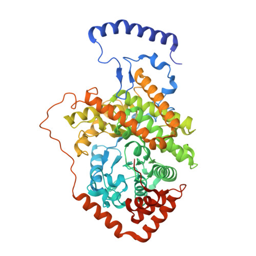
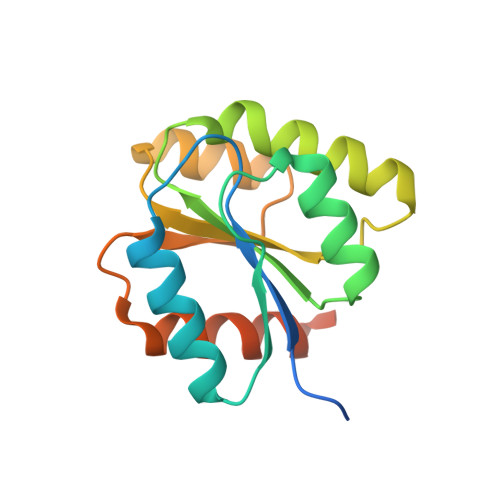
    4R3U

  • PubMed Abstract: 

    Bacterial coenzyme B12-dependent 2-hydroxyisobutyryl-CoA mutase (HCM) is a radical enzyme catalyzing the stereospecific interconversion of (S)-3-hydroxybutyryl- and 2-hydroxyisobutyryl-CoA. It consists of two subunits, HcmA and HcmB. To characterize the determinants of substrate specificity, we have analyzed the crystal structure of HCM from Aquincola tertiaricarbonis in complex with coenzyme B12 and the substrates (S)-3-hydroxybutyryl- and 2-hydroxyisobutyryl-CoA in alternative binding. When compared with the well studied structure of bacterial and mitochondrial B12-dependent methylmalonyl-CoA mutase (MCM), HCM has a highly conserved domain architecture. However, inspection of the substrate binding site identified amino acid residues not present in MCM, namely HcmA Ile(A90) and Asp(A117). Asp(A117) determines the orientation of the hydroxyl group of the acyl-CoA esters by H-bond formation, thus determining stereospecificity of catalysis. Accordingly, HcmA D117A and D117V mutations resulted in significantly increased activity toward (R)-3-hydroxybutyryl-CoA. Besides interconversion of hydroxylated acyl-CoA esters, wild-type HCM as well as HcmA I90V and I90A mutant enzymes could also isomerize pivalyl- and isovaleryl-CoA, albeit at >10 times lower rates than the favorite substrate (S)-3-hydroxybutyryl-CoA. The nonconservative mutation HcmA D117V, however, resulted in an enzyme showing high activity toward pivalyl-CoA. Structural requirements for binding and isomerization of highly branched acyl-CoA substrates such as 2-hydroxyisobutyryl- and pivalyl-CoA, possessing tertiary and quaternary carbon atoms, respectively, are discussed.


  • Organizational Affiliation

    From the Department of Environmental Microbiology, Helmholtz Centre for Environmental Research (UFZ), 04318 Leipzig and.


Macromolecules
Find similar proteins by:  (by identity cutoff)  |  3D Structure
Entity ID: 1
MoleculeChains Sequence LengthOrganismDetailsImage
2-hydroxyisobutyryl-CoA mutase large subunit
A, B
584Aquincola tertiaricarbonisMutation(s): 0 
Gene Names: hcmA
EC: 5.4.99
UniProt
Find proteins for I3VE77 (Aquincola tertiaricarbonis)
Explore I3VE77 
Go to UniProtKB:  I3VE77
Entity Groups  
Sequence Clusters30% Identity50% Identity70% Identity90% Identity95% Identity100% Identity
UniProt GroupI3VE77
Sequence Annotations
Expand
  • Reference Sequence
Find similar proteins by:  (by identity cutoff)  |  3D Structure
Entity ID: 2
MoleculeChains Sequence LengthOrganismDetailsImage
2-hydroxyisobutyryl-CoA mutase small subunit
C, D
158Aquincola tertiaricarbonisMutation(s): 0 
Gene Names: hcmB
UniProt
Find proteins for I3VE74 (Aquincola tertiaricarbonis)
Explore I3VE74 
Go to UniProtKB:  I3VE74
Entity Groups  
Sequence Clusters30% Identity50% Identity70% Identity90% Identity95% Identity100% Identity
UniProt GroupI3VE74
Sequence Annotations
Expand
  • Reference Sequence
Small Molecules
Ligands 4 Unique
IDChains Name / Formula / InChI Key2D Diagram3D Interactions
B12
Query on B12

Download Ideal Coordinates CCD File 
J [auth C],
K [auth D]
COBALAMIN
C62 H89 Co N13 O14 P
LKVIQTCSMMVGFU-DWSMJLPVSA-N
3KK
Query on 3KK

Download Ideal Coordinates CCD File 
F [auth A],
I [auth B]
S-{(3R,5R,9R)-1-[(2R,3S,4R,5R)-5-(6-amino-9H-purin-9-yl)-4-hydroxy-3-(phosphonooxy)tetrahydrofuran-2-yl]-3,5,9-trihydroxy-8,8-dimethyl-3,5-dioxido-10,14-dioxo-2,4,6-trioxa-11,15-diaza-3lambda~5~,5lambda~5~-diphosphaheptadecan-17-yl} 2-hydroxy-2-methylpropanethioate
C25 H42 N7 O18 P3 S
FFVUICCDNWZCRC-ZSJPKINUSA-N
3HC
Query on 3HC

Download Ideal Coordinates CCD File 
E [auth A],
H [auth B]
3-HYDROXYBUTANOYL-COENZYME A
C25 H42 N7 O18 P3 S
QHHKKMYHDBRONY-VKBDFPRVSA-N
5AD
Query on 5AD

Download Ideal Coordinates CCD File 
G [auth A]5'-DEOXYADENOSINE
C10 H13 N5 O3
XGYIMTFOTBMPFP-KQYNXXCUSA-N
Experimental Data & Validation

Experimental Data

  • Method: X-RAY DIFFRACTION
  • Resolution: 2.50 Å
  • R-Value Free: 0.217 
  • R-Value Work: 0.168 
  • R-Value Observed: 0.170 
  • Space Group: C 1 2 1
Unit Cell:
Length ( Å )Angle ( ˚ )
a = 69.03α = 90
b = 119.56β = 91.23
c = 173.94γ = 90
Software Package:
Software NamePurpose
MOLREPphasing
BUSTERrefinement
XDSdata reduction
SCALAdata scaling

Structure Validation

View Full Validation Report


Ligand Structure Quality Assessment 


Entry History 

Deposition Data

Revision History  (Full details and data files)

  • Version 1.0: 2015-03-11
    Type: Initial release
  • Version 1.1: 2015-06-17
    Changes: Database references
  • Version 1.2: 2024-02-28
    Changes: Data collection, Database references, Derived calculations