4R0D

Crystal structure of a eukaryotic group II intron lariat


Experimental Data Snapshot

  • Method: X-RAY DIFFRACTION
  • Resolution: 3.68 Å
  • R-Value Free: 0.274 
  • R-Value Work: 0.239 
  • R-Value Observed: 0.241 

wwPDB Validation   3D Report Full Report


Ligand Structure Quality Assessment 


This is version 1.5 of the entry. See complete history

Re-refinement Note

A newer entry is available that reflects an alternative modeling of the original data: 6CIH


Literature

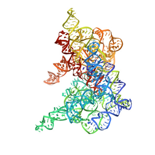
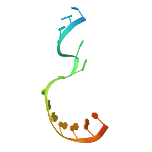
Crystal structure of a eukaryotic group II intron lariat.

Robart, A.R.Chan, R.T.Peters, J.K.Rajashankar, K.R.Toor, N.

(2014) Nature 514: 193-197

  • DOI: https://doi.org/10.1038/nature13790
  • Primary Citation of Related Structures:  
    4R0D

  • PubMed Abstract: 

    The formation of branched lariat RNA is an evolutionarily conserved feature of splicing reactions for both group II and spliceosomal introns. The lariat is important for the fidelity of 5' splice-site selection and consists of a 2'-5' phosphodiester bond between a bulged adenosine and the 5' end of the intron. To gain insight into this ubiquitous intramolecular linkage, we determined the crystal structure of a eukaryotic group IIB intron in the lariat form at 3.7 Å. This revealed that two tandem tetraloop-receptor interactions, η-η' and π-π', place domain VI in the core to position the lariat bond in the post-catalytic state. On the basis of structural and biochemical data, we propose that π-π' is a dynamic interaction that mediates the transition between the two steps of splicing, with η-η' serving an ancillary role. The structure also reveals a four-magnesium-ion cluster involved in both catalysis and positioning of the 5' end. Given the evolutionary relationship between group II and nuclear introns, it is likely that this active site configuration exists in the spliceosome as well.


  • Organizational Affiliation

    Department of Chemistry and Biochemistry, University of California, San Diego, La Jolla, California 92093, USA.


Macromolecules
Find similar nucleic acids by:  (by identity cutoff)  |  3D Structure
Entity ID: 1
MoleculeChains LengthOrganismImage
GROUP IIB INTRON LARIAT622Pylaiella littoralis
Sequence Annotations
Expand
  • Reference Sequence

Find similar nucleic acids by:  Sequence   |   3D Structure  

Entity ID: 2
MoleculeChains LengthOrganismImage
LIGATED EXONS20Pylaiella littoralis
Sequence Annotations
Expand
  • Reference Sequence
Small Molecules
Ligands 4 Unique
IDChains Name / Formula / InChI Key2D Diagram3D Interactions
IRI
Query on IRI

Download Ideal Coordinates CCD File 
AA [auth A]
BA [auth A]
CA [auth A]
DA [auth A]
E [auth A]
AA [auth A],
BA [auth A],
CA [auth A],
DA [auth A],
E [auth A],
EA [auth A],
F [auth A],
FA [auth A],
G [auth A],
GA [auth A],
H [auth A],
HA [auth A],
I [auth A],
IA [auth A],
J [auth A],
JA [auth A],
K [auth A],
KA [auth A],
L [auth A],
LA [auth A],
M [auth A],
MA [auth A],
N [auth A],
NA [auth A],
O [auth A],
OA [auth A],
P [auth A],
PA [auth A],
Q [auth A],
R [auth A],
S [auth A],
T [auth A],
U [auth A],
V [auth A],
W [auth A],
X [auth A],
Y [auth A],
Z [auth A]
IRIDIUM HEXAMMINE ION
H18 Ir N6
CGMAOQLDNKCXGK-UHFFFAOYSA-N
SPM
Query on SPM

Download Ideal Coordinates CCD File 
C [auth A],
D [auth A]
SPERMINE
C10 H26 N4
PFNFFQXMRSDOHW-UHFFFAOYSA-N
MG
Query on MG

Download Ideal Coordinates CCD File 
AB [auth A]
AC [auth A]
AD [auth A]
BB [auth A]
BC [auth A]
AB [auth A],
AC [auth A],
AD [auth A],
BB [auth A],
BC [auth A],
BD [auth A],
CB [auth A],
CC [auth A],
CD [auth A],
DB [auth A],
DC [auth A],
DD [auth A],
EB [auth A],
EC [auth A],
ED [auth A],
FB [auth A],
FC [auth A],
FD [auth A],
GB [auth A],
GC [auth A],
GD [auth A],
HB [auth A],
HC [auth A],
HD [auth A],
IB [auth A],
IC [auth A],
ID [auth A],
JB [auth A],
JC [auth A],
JD [auth A],
JE [auth B],
KB [auth A],
KC [auth A],
KD [auth A],
KE [auth B],
LB [auth A],
LC [auth A],
LD [auth A],
MB [auth A],
MC [auth A],
MD [auth A],
NB [auth A],
NC [auth A],
ND [auth A],
OB [auth A],
OC [auth A],
OD [auth A],
PB [auth A],
PC [auth A],
PD [auth A],
QA [auth A],
QB [auth A],
QC [auth A],
QD [auth A],
RA [auth A],
RB [auth A],
RC [auth A],
RD [auth A],
SA [auth A],
SB [auth A],
SC [auth A],
SD [auth A],
TA [auth A],
TB [auth A],
TC [auth A],
UA [auth A],
UB [auth A],
UC [auth A],
VA [auth A],
VB [auth A],
VC [auth A],
WA [auth A],
WB [auth A],
WC [auth A],
XA [auth A],
XB [auth A],
XC [auth A],
YA [auth A],
YB [auth A],
YC [auth A],
ZA [auth A],
ZB [auth A],
ZC [auth A]
MAGNESIUM ION
Mg
JLVVSXFLKOJNIY-UHFFFAOYSA-N
NA
Query on NA

Download Ideal Coordinates CCD File 
AE [auth A]
BE [auth A]
CE [auth A]
DE [auth A]
EE [auth A]
AE [auth A],
BE [auth A],
CE [auth A],
DE [auth A],
EE [auth A],
FE [auth A],
GE [auth A],
HE [auth A],
IE [auth A],
TD [auth A],
UD [auth A],
VD [auth A],
WD [auth A],
XD [auth A],
YD [auth A],
ZD [auth A]
SODIUM ION
Na
FKNQFGJONOIPTF-UHFFFAOYSA-N
Experimental Data & Validation

Experimental Data

  • Method: X-RAY DIFFRACTION
  • Resolution: 3.68 Å
  • R-Value Free: 0.274 
  • R-Value Work: 0.239 
  • R-Value Observed: 0.241 
  • Space Group: C 2 2 21
Unit Cell:
Length ( Å )Angle ( ˚ )
a = 163.639α = 90
b = 255.361β = 90
c = 136.786γ = 90
Software Package:
Software NamePurpose
HKL-2000data collection
SHELXmodel building
PHENIXrefinement
HKL-2000data reduction
HKL-2000data scaling
SHELXphasing

Structure Validation

View Full Validation Report


Ligand Structure Quality Assessment 


Entry History 

Deposition Data

Revision History  (Full details and data files)

  • Version 1.0: 2014-10-01
    Type: Initial release
  • Version 1.1: 2014-10-08
    Changes: Database references
  • Version 1.2: 2014-10-15
    Changes: Database references
  • Version 1.3: 2014-10-29
    Changes: Source and taxonomy
  • Version 1.4: 2016-05-25
    Changes: Source and taxonomy
  • Version 1.5: 2024-02-28
    Changes: Data collection, Database references, Derived calculations