4QAM

Crystal Structure of the RPGR RCC1-like domain in complex with the RPGR-interacting domain of RPGRIP1


Experimental Data Snapshot

  • Method: X-RAY DIFFRACTION
  • Resolution: 1.83 Å
  • R-Value Free: 0.197 
  • R-Value Work: 0.162 
  • R-Value Observed: 0.164 

wwPDB Validation   3D Report Full Report


This is version 1.3 of the entry. See complete history


Literature

C2 domains as protein-protein interaction modules in the ciliary transition zone.

Remans, K.Burger, M.Vetter, I.R.Wittinghofer, A.

(2014) Cell Rep 8: 1-9

  • DOI: https://doi.org/10.1016/j.celrep.2014.05.049
  • Primary Citation of Related Structures:  
    4QAM

  • PubMed Abstract: 

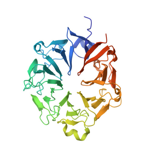
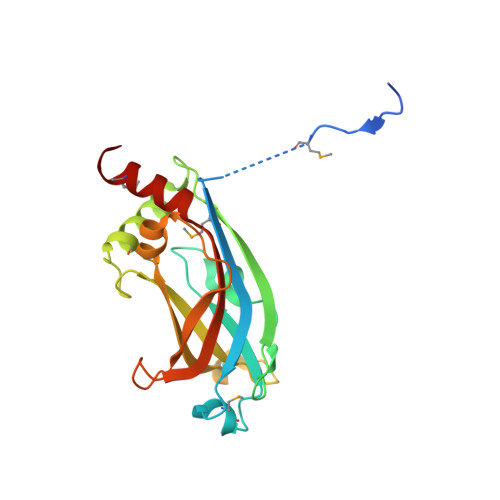
    RPGR-interacting protein 1 (RPGRIP1) is mutated in the eye disease Leber congenital amaurosis (LCA) and its structural homolog, RPGRIP1-like (RPGRIP1L), is mutated in many different ciliopathies. Both are multidomain proteins that are predicted to interact with retinitis pigmentosa G-protein regulator (RPGR). RPGR is mutated in X-linked retinitis pigmentosa and is located in photoreceptors and primary cilia. We solved the crystal structure of the complex between the RPGR-interacting domain (RID) of RPGRIP1 and RPGR and demonstrate that RPGRIP1L binds to RPGR similarly. RPGRIP1 binding to RPGR affects the interaction with PDEδ, the cargo shuttling factor for prenylated ciliary proteins. RPGRIP1-RID is a C2 domain with a canonical β sandwich structure that does not bind Ca(2+) and/or phospholipids and thus constitutes a unique type of protein-protein interaction module. Judging from the large number of C2 domains in most of the ciliary transition zone proteins identified thus far, the structure presented here seems to constitute a cilia-specific module that is present in multiprotein transition zone complexes.


  • Organizational Affiliation

    Max Planck Institute of Molecular Physiology, Otto-Hahn-Strasse 11, 44227 Dortmund, Germany.


Macromolecules
Find similar proteins by:  (by identity cutoff)  |  3D Structure
Entity ID: 1
MoleculeChains Sequence LengthOrganismDetailsImage
X-linked retinitis pigmentosa GTPase regulator400Homo sapiensMutation(s): 1 
Gene Names: RP3RPGRXLRP3
UniProt & NIH Common Fund Data Resources
Find proteins for Q92834 (Homo sapiens)
Explore Q92834 
Go to UniProtKB:  Q92834
PHAROS:  Q92834
GTEx:  ENSG00000156313 
Entity Groups  
Sequence Clusters30% Identity50% Identity70% Identity90% Identity95% Identity100% Identity
UniProt GroupQ92834
Sequence Annotations
Expand
  • Reference Sequence
Find similar proteins by:  (by identity cutoff)  |  3D Structure
Entity ID: 2
MoleculeChains Sequence LengthOrganismDetailsImage
X-linked retinitis pigmentosa GTPase regulator-interacting protein 1201Homo sapiensMutation(s): 0 
Gene Names: RPGRIP1
UniProt & NIH Common Fund Data Resources
Find proteins for Q96KN7 (Homo sapiens)
Explore Q96KN7 
Go to UniProtKB:  Q96KN7
PHAROS:  Q96KN7
GTEx:  ENSG00000092200 
Entity Groups  
Sequence Clusters30% Identity50% Identity70% Identity90% Identity95% Identity100% Identity
UniProt GroupQ96KN7
Sequence Annotations
Expand
  • Reference Sequence
Small Molecules
Modified Residues  1 Unique
IDChains TypeFormula2D DiagramParent
MSE
Query on MSE
B
L-PEPTIDE LINKINGC5 H11 N O2 SeMET
Experimental Data & Validation

Experimental Data

  • Method: X-RAY DIFFRACTION
  • Resolution: 1.83 Å
  • R-Value Free: 0.197 
  • R-Value Work: 0.162 
  • R-Value Observed: 0.164 
  • Space Group: P 21 21 21
Unit Cell:
Length ( Å )Angle ( ˚ )
a = 63.12α = 90
b = 63.8β = 90
c = 161.44γ = 90
Software Package:
Software NamePurpose
XSCALEdata scaling
REFMACrefinement
PDB_EXTRACTdata extraction
PHENIXphasing

Structure Validation

View Full Validation Report


Entry History 

Deposition Data

Revision History  (Full details and data files)

  • Version 1.0: 2014-07-30
    Type: Initial release
  • Version 1.1: 2014-11-19
    Changes: Experimental preparation
  • Version 1.2: 2023-09-20
    Changes: Data collection, Database references, Derived calculations, Refinement description
  • Version 1.3: 2023-12-06
    Changes: Data collection