


Crystal Structure of a Beta-1,3-glucanase from Mycobacterium marinum
Dranow, D.M., Davies, D.R., Edwards, T.E., Lorimer, D., Seattle Structural Genomics Center for Infectious DiseaseTo be published.
Experimental Data Snapshot
wwPDB Validation 3D Report Full Report

Entity ID: 1 | |||||
|---|---|---|---|---|---|
| Molecule | Chains | Sequence Length | Organism | Details | Image |
| Beta-1,3-glucanase | 257 | Mycobacterium marinum M | Mutation(s): 0 Gene Names: MMAR_2902 | 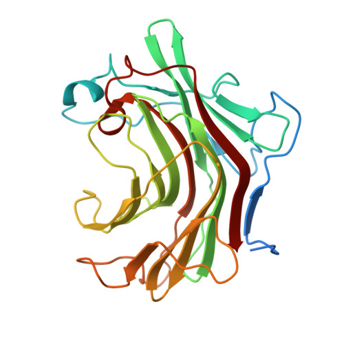 | |
UniProt | |||||
Find proteins for B2HE62 (Mycobacterium marinum (strain ATCC BAA-535 / M)) Explore B2HE62 Go to UniProtKB: B2HE62 | |||||
Entity Groups | |||||
| Sequence Clusters | 30% Identity50% Identity70% Identity90% Identity95% Identity100% Identity | ||||
| UniProt Group | B2HE62 | ||||
Sequence AnnotationsExpand | |||||
| |||||
| Ligands 3 Unique | |||||
|---|---|---|---|---|---|
| ID | Chains | Name / Formula / InChI Key | 2D Diagram | 3D Interactions | |
| BTB Query on BTB | E [auth A], H [auth B] | 2-[BIS-(2-HYDROXY-ETHYL)-AMINO]-2-HYDROXYMETHYL-PROPANE-1,3-DIOL C8 H19 N O5 OWMVSZAMULFTJU-UHFFFAOYSA-N |  | ||
| EDO Query on EDO | F [auth A] | 1,2-ETHANEDIOL C2 H6 O2 LYCAIKOWRPUZTN-UHFFFAOYSA-N |  | ||
| MG Query on MG | C [auth A], D [auth A], G [auth B] | MAGNESIUM ION Mg JLVVSXFLKOJNIY-UHFFFAOYSA-N |  | ||
| Length ( Å ) | Angle ( ˚ ) |
|---|---|
| a = 45.12 | α = 109.45 |
| b = 46.84 | β = 95.63 |
| c = 51.84 | γ = 99.79 |
| Software Name | Purpose |
|---|---|
| XSCALE | data scaling |
| REFMAC | refinement |
| PDB_EXTRACT | data extraction |
| BALBES | phasing |
RCSB PDB (citation) is hosted by
RCSB PDB is a member of the

