


The structure of Japanese Marasmius oreades lectin at 1.40 Angstroms resolution.
Noma, Y., Shimokawa, M., Maeganeku, C., Motoshima, H., Watanabe, K., Minami, Y., Yagi, F.To be published.
Experimental Data Snapshot
wwPDB Validation 3D Report Full Report

Entity ID: 1 | |||||
|---|---|---|---|---|---|
| Molecule | Chains | Sequence Length | Organism | Details | Image |
| Mannose recognizing lectin | 120 | Marasmius oreades | Mutation(s): 0 Gene Names: Mlec1 | 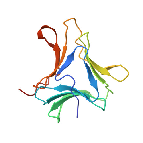 | |
UniProt | |||||
Find proteins for I7H471 (Marasmius oreades) Explore I7H471 Go to UniProtKB: I7H471 | |||||
Entity Groups | |||||
| Sequence Clusters | 30% Identity50% Identity70% Identity90% Identity95% Identity100% Identity | ||||
| UniProt Group | I7H471 | ||||
Sequence AnnotationsExpand | |||||
| |||||
| Ligands 1 Unique | |||||
|---|---|---|---|---|---|
| ID | Chains | Name / Formula / InChI Key | 2D Diagram | 3D Interactions | |
| SO4 Query on SO4 | B [auth A], C [auth A], D [auth A] | SULFATE ION O4 S QAOWNCQODCNURD-UHFFFAOYSA-L |  | ||
| Length ( Å ) | Angle ( ˚ ) |
|---|---|
| a = 57.395 | α = 90 |
| b = 57.395 | β = 90 |
| c = 57.944 | γ = 120 |
| Software Name | Purpose |
|---|---|
| REFMAC | refinement |
RCSB PDB (citation) is hosted by
RCSB PDB is a member of the

