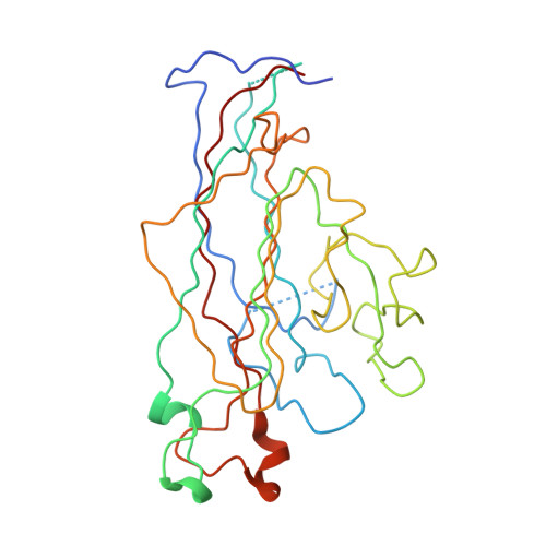
Structure analysis of the major capsid proteins of human polyomaviruses 6 and 7 reveals an obstructed sialic Acid binding site.
Stroh, L.J., Neu, U., Blaum, B.S., Buch, M.H., Garcea, R.L., Stehle, T.(2014) J Virol 88: 10831-10839
- PubMed: 25008942
- DOI: https://doi.org/10.1128/JVI.01084-14
- Primary Citation of Related Structures:
4PCG, 4PCH - PubMed Abstract:
Human polyomavirus 6 (HPyV6) and HPyV7 are commonly found on human skin. We have determined the X-ray structures of their major capsid protein, VP1, at resolutions of 1.8 and 1.7 Å, respectively. In polyomaviruses, VP1 commonly determines antigenicity as well as cell-surface receptor specificity, and the protein is therefore linked to attachment, tropism, and ultimately, viral pathogenicity. The structures of HPyV6 and HPyV7 VP1 reveal uniquely elongated loops that cover the bulk of the outer virion surfaces, obstructing a groove that binds sialylated glycan receptors in many other polyomaviruses. In support of this structural observation, interactions of VP1 with α2,3- and α2,6-linked sialic acids could not be detected in solution by nuclear magnetic resonance spectroscopy. Single-cell binding studies indicate that sialylated glycans are likely not required for initial attachment to cultured human cells. Our findings establish distinct antigenic properties of HPyV6 and HPyV7 capsids and indicate that these two viruses engage nonsialylated receptors. Eleven new human polyomaviruses, including the skin viruses HPyV6 and HPyV7, have been identified during the last decade. In contrast to better-studied polyomaviruses, the routes of infection, cell tropism, and entry pathways of many of these new viruses remain largely mysterious. Our high-resolution X-ray structures of major capsid proteins VP1 from HPyV6 and from HPyV7 reveal critical differences in surface morphology from those of all other known polyomavirus structures. A groove that engages specific sialic acid-containing glycan receptors in related polyomaviruses is obstructed, and VP1 of HPyV6 and HPyV7 does not interact with sialylated compounds in solution or on cultured human cells. A comprehensive comparison with other structurally characterized polyomavirus VP1 proteins enhances our understanding of molecular determinants that underlie receptor specificity, antigenicity, and, ultimately, pathogenicity within the polyomavirus family and highlight the need for structure-based analysis to better define phylogenetic relationships within the growing polyomavirus family and perhaps also for other viruses.
Organizational Affiliation:
Interfaculty Institute of Biochemistry, University of Tübingen, Tübingen, Germany.