4P6T

Crystal Structure of tyrosinase from Bacillus megaterium with p-tyrosol in the active site


Experimental Data Snapshot

  • Method: X-RAY DIFFRACTION
  • Resolution: 2.50 Å
  • R-Value Free: 0.261 
  • R-Value Work: 0.208 

wwPDB Validation   3D Report Full Report


This is version 1.3 of the entry. See complete history


Literature

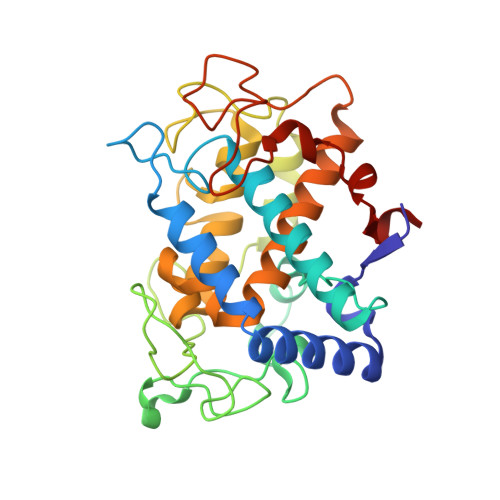
Determination of tyrosinase substrate-binding modes reveals mechanistic differences between type-3 copper proteins.

Goldfeder, M.Kanteev, M.Isaschar-Ovdat, S.Adir, N.Fishman, A.

(2014) Nat Commun 5: 4505-4505

  • DOI: https://doi.org/10.1038/ncomms5505
  • Primary Citation of Related Structures:  
    4P6R, 4P6S, 4P6T

  • PubMed Abstract: 

    Tyrosinase is responsible for the two initial enzymatic steps in the conversion of tyrosine to melanin. Many tyrosinase mutations are the leading cause of albinism in humans, and it is a prominent biotechnology and pharmaceutical industry target. Here we present crystal structures that show that both monophenol hydroxylation and diphenol oxidation occur at the same site. It is suggested that concurrent presence of a phenylalanine above the active site and a restricting thioether bond on the histidine coordinating CuA prevent hydroxylation of monophenols by catechol oxidases. Furthermore, a conserved water molecule activated by E195 and N205 is proposed to mediate deprotonation of the monophenol at the active site. Overall, the structures reveal precise steps in the enzymatic catalytic cycle as well as differences between tyrosinases and other type-3 copper enzymes.


  • Organizational Affiliation

    1] Department of Biotechnology and Food Engineering, Technion-Israel Institute of Technology, Haifa 32000, Israel [2].


Macromolecules
Find similar proteins by:  (by identity cutoff)  |  3D Structure
Entity ID: 1
MoleculeChains Sequence LengthOrganismDetailsImage
Tyrosinase
A, B
287Priestia megateriumMutation(s): 0 
EC: 1.14.18.1
UniProt
Find proteins for B2ZB02 (Priestia megaterium)
Explore B2ZB02 
Go to UniProtKB:  B2ZB02
Entity Groups  
Sequence Clusters30% Identity50% Identity70% Identity90% Identity95% Identity100% Identity
UniProt GroupB2ZB02
Sequence Annotations
Expand
  • Reference Sequence
Experimental Data & Validation

Experimental Data

  • Method: X-RAY DIFFRACTION
  • Resolution: 2.50 Å
  • R-Value Free: 0.261 
  • R-Value Work: 0.208 
  • Space Group: P 1 21 1
Unit Cell:
Length ( Å )Angle ( ˚ )
a = 47.69α = 90
b = 78.09β = 105.96
c = 84.28γ = 90
Software Package:
Software NamePurpose
MOSFLMdata reduction
SCALAdata scaling
PHENIXrefinement
PDB_EXTRACTdata extraction
PHASERphasing

Structure Validation

View Full Validation Report


Entry History & Funding Information

Deposition Data


Funding OrganizationLocationGrant Number
Israel Science FoundationIsrael193/11

Revision History  (Full details and data files)

  • Version 1.0: 2014-07-30
    Type: Initial release
  • Version 1.1: 2014-08-06
    Changes: Database references
  • Version 1.2: 2017-11-22
    Changes: Advisory, Derived calculations, Other, Refinement description, Source and taxonomy
  • Version 1.3: 2023-12-27
    Changes: Data collection, Database references, Derived calculations, Refinement description