


Crystal Structure of a Thioredoxin Mutant Displays a Dynamic N-terminal Loop Surrounding an S-nitrosation Site
The, J., Weichsel, A., Montfort, W.R.To be published.
Experimental Data Snapshot
Starting Model: experimental
View more details
wwPDB Validation 3D Report Full Report

Entity ID: 1 | |||||
|---|---|---|---|---|---|
| Molecule | Chains | Sequence Length | Organism | Details | Image |
| Thioredoxin | 105 | Homo sapiens | Mutation(s): 3 Gene Names: TXN, TRDX, TRX, TRX1 | 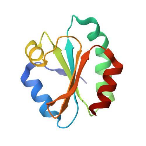 | |
UniProt & NIH Common Fund Data Resources | |||||
Find proteins for P10599 (Homo sapiens) Explore P10599 Go to UniProtKB: P10599 | |||||
PHAROS: P10599 GTEx: ENSG00000136810 | |||||
Entity Groups | |||||
| Sequence Clusters | 30% Identity50% Identity70% Identity90% Identity95% Identity100% Identity | ||||
| UniProt Group | P10599 | ||||
Sequence AnnotationsExpand | |||||
| |||||
| Length ( Å ) | Angle ( ˚ ) |
|---|---|
| a = 35.269 | α = 90 |
| b = 50.182 | β = 94.08 |
| c = 51.066 | γ = 90 |
| Software Name | Purpose |
|---|---|
| XDS | data scaling |
| MOLREP | phasing |
| REFMAC | refinement |
| HKL-2000 | data reduction |
| SCALA | data scaling |
RCSB PDB (citation) is hosted by
RCSB PDB is a member of the

