4O26

Crystal structure of the TRBD domain of TERT and the CR4/5 of TR


Experimental Data Snapshot

  • Method: X-RAY DIFFRACTION
  • Resolution: 3.00 Å
  • R-Value Free: 0.261 
  • R-Value Work: 0.221 
  • R-Value Observed: 0.223 

wwPDB Validation   3D Report Full Report


This is version 1.2 of the entry. See complete history


Literature

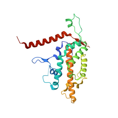
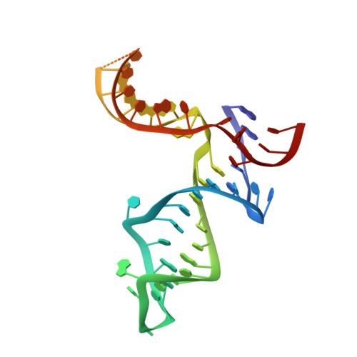
Structural basis for protein-RNA recognition in telomerase.

Huang, J.Brown, A.F.Wu, J.Xue, J.Bley, C.J.Rand, D.P.Wu, L.Zhang, R.Chen, J.J.Lei, M.

(2014) Nat Struct Mol Biol 21: 507-512

  • DOI: https://doi.org/10.1038/nsmb.2819
  • Primary Citation of Related Structures:  
    4O26

  • PubMed Abstract: 

    Telomerase is a large ribonucleoprotein complex minimally composed of a catalytic telomerase reverse transcriptase (TERT) and an RNA component (TR) that provides the template for telomeric DNA synthesis. However, it remains unclear how TERT and TR assemble into a functional telomerase. Here we report the crystal structure of the conserved regions 4 and 5 (CR4/5) of TR in complex with the TR-binding domain (TRBD) of TERT from the teleost fish Oryzias latipes. The structure shows that CR4/5 adopts an L-shaped three-way-junction conformation with its two arms clamping onto TRBD. Both the sequence and conformation of CR4/5 are required for the interaction. Our structural and mutational analyses suggest that the observed CR4/5-TRBD recognition is common to most eukaryotes, and CR4/5 in vertebrate TR might have a similar role in telomerase regulation as that of stem-loop IV in Tetrahymena TR.


  • Organizational Affiliation

    1] National Center for Protein Science Shanghai, State Key Laboratory of Molecular Biology, Institute of Biochemistry and Cell Biology, Shanghai Institutes for Biological Sciences, Chinese Academy of Sciences, Shanghai, China. [2] Howard Hughes Medical Institute, University of Michigan Medical School, Ann Arbor, Michigan, USA. [3] Department of Biological Chemistry, University of Michigan Medical School, Ann Arbor, Michigan, USA.


Macromolecules

Find similar proteins by:  (by identity cutoff)  |  3D Structure
Entity ID: 1
MoleculeChains Sequence LengthOrganismDetailsImage
Telomerase reverse transcriptase
A, B
257Oryzias latipesMutation(s): 0 
Gene Names: TERT
UniProt
Find proteins for Q1PS67 (Oryzias latipes)
Explore Q1PS67 
Go to UniProtKB:  Q1PS67
Entity Groups  
Sequence Clusters30% Identity50% Identity70% Identity90% Identity95% Identity100% Identity
UniProt GroupQ1PS67
Sequence Annotations
Expand
  • Reference Sequence
Find similar nucleic acids by:  (by identity cutoff)  |  3D Structure
Entity ID: 2
MoleculeChains LengthOrganismImage
Telomerase TRC [auth E],
D [auth F]
51Oryzias latipes
Sequence Annotations
Expand
  • Reference Sequence
Small Molecules
Ligands 1 Unique
IDChains Name / Formula / InChI Key2D Diagram3D Interactions
SO4
Query on SO4

Download Ideal Coordinates CCD File 
E [auth A]
F [auth A]
G [auth A]
H [auth A]
I [auth A]
E [auth A],
F [auth A],
G [auth A],
H [auth A],
I [auth A],
J [auth A],
K [auth A],
L [auth A],
M [auth B],
N [auth B],
O [auth B],
P [auth B]
SULFATE ION
O4 S
QAOWNCQODCNURD-UHFFFAOYSA-L
Experimental Data & Validation

Experimental Data

  • Method: X-RAY DIFFRACTION
  • Resolution: 3.00 Å
  • R-Value Free: 0.261 
  • R-Value Work: 0.221 
  • R-Value Observed: 0.223 
  • Space Group: P 62 2 2
Unit Cell:
Length ( Å )Angle ( ˚ )
a = 118.092α = 90
b = 118.092β = 90
c = 358.021γ = 120
Software Package:
Software NamePurpose
PHENIXrefinement
REFMACrefinement
PDB_EXTRACTdata extraction
SCALEPACKdata scaling

Structure Validation

View Full Validation Report


Entry History 

Deposition Data

Revision History  (Full details and data files)

  • Version 1.0: 2014-05-07
    Type: Initial release
  • Version 1.1: 2014-06-18
    Changes: Database references
  • Version 1.2: 2019-07-17
    Changes: Data collection, Refinement description