4NT5

Crystal structure of human von Willebrand factor CTCK domain


Experimental Data Snapshot

  • Method: X-RAY DIFFRACTION
  • Resolution: 3.28 Å
  • R-Value Free: 0.248 
  • R-Value Work: 0.224 
  • R-Value Observed: 0.226 

wwPDB Validation   3D Report Full Report


Ligand Structure Quality Assessment 


This is version 1.4 of the entry. See complete history


Literature

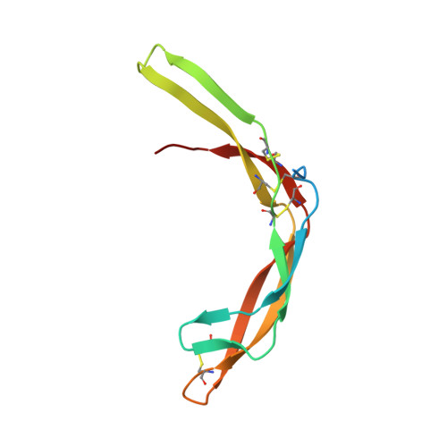
Highly reinforced structure of a C-terminal dimerization domain in von Willebrand factor.

Zhou, Y.F.Springer, T.A.

(2014) Blood 123: 1785-1793

  • DOI: https://doi.org/10.1182/blood-2013-11-523639
  • Primary Citation of Related Structures:  
    4NT5

  • PubMed Abstract: 

    The C-terminal cystine knot (CK) (CTCK) domain in von Willebrand factor (VWF) mediates dimerization of proVWF in the endoplasmic reticulum and is essential for long multimers required for hemostatic function. The CTCK dimer crystal structure reveals highly elongated monomers with 2 β-ribbons and 4 intra-chain disulfides, including 3 in the CK. Dimerization buries an extensive interface of 1500 Å(2) corresponding to 32% of the surface of each monomer and forms a super β-sheet and 3 inter-chain disulfides. The shape, dimensions, and N-terminal connections of the crystal structure agree perfectly with previous electron microscopic images of VWF dimeric bouquets with the CTCK dimer forming a down-curved base. The dimer interface is suited to resist hydrodynamic force and disulfide reduction. CKs in each monomer flank the 3 inter-chain disulfides, and their presence in β-structures with dense backbone hydrogen bonds creates a rigid, highly crosslinked interface. The structure reveals the basis for von Willebrand disease phenotypes and the fold and disulfide linkages for CTCK domains in diverse protein families involved in barrier function, eye and inner ear development, insect coagulation and innate immunity, axon guidance, and signaling in extracellular matrices.


  • Organizational Affiliation

    Program in Cellular and Molecular Medicine, Children's Hospital Boston and Department of Biological Chemistry and Molecular Pharmacology, Harvard Medical School, Boston, MA.


Macromolecules
Find similar proteins by:  (by identity cutoff)  |  3D Structure
Entity ID: 1
MoleculeChains Sequence LengthOrganismDetailsImage
von Willebrand factor107Homo sapiensMutation(s): 0 
Gene Names: F8VWFVWF
UniProt & NIH Common Fund Data Resources
Find proteins for P04275 (Homo sapiens)
Explore P04275 
Go to UniProtKB:  P04275
PHAROS:  P04275
GTEx:  ENSG00000110799 
Entity Groups  
Sequence Clusters30% Identity50% Identity70% Identity90% Identity95% Identity100% Identity
UniProt GroupP04275
Sequence Annotations
Expand
  • Reference Sequence
Experimental Data & Validation

Experimental Data

  • Method: X-RAY DIFFRACTION
  • Resolution: 3.28 Å
  • R-Value Free: 0.248 
  • R-Value Work: 0.224 
  • R-Value Observed: 0.226 
  • Space Group: P 43 3 2
Unit Cell:
Length ( Å )Angle ( ˚ )
a = 135.34α = 90
b = 135.34β = 90
c = 135.34γ = 90
Software Package:
Software NamePurpose
PHASERphasing
PHENIXrefinement
XDSdata reduction
XSCALEdata scaling

Structure Validation

View Full Validation Report


Ligand Structure Quality Assessment 


Entry History 

Deposition Data

Revision History  (Full details and data files)

  • Version 1.0: 2014-01-01
    Type: Initial release
  • Version 1.1: 2014-04-02
    Changes: Database references
  • Version 1.2: 2014-04-09
    Changes: Derived calculations
  • Version 1.3: 2020-07-29
    Type: Remediation
    Reason: Carbohydrate remediation
    Changes: Data collection, Database references, Derived calculations, Structure summary
  • Version 1.4: 2024-01-10
    Changes: Advisory, Data collection, Database references, Derived calculations, Structure summary