N-terminal protein modification by substrate-activated reverse proteolysis.
Liebscher, S., Schopfel, M., Aumuller, T., Sharkhuukhen, A., Pech, A., Hoss, E., Parthier, C., Jahreis, G., Stubbs, M.T., Bordusa, F.(2014) Angew Chem Int Ed Engl 53: 3024-3028
- PubMed: 24520050
- DOI: https://doi.org/10.1002/anie.201307736
- Primary Citation of Related Structures:
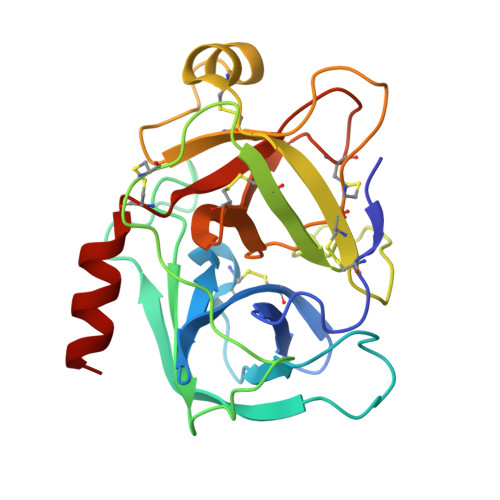
4NIV, 4NIW, 4NIX, 4NIY - PubMed Abstract:
Although site-specific incorporation of artificial functionalities into proteins is an important tool in both basic and applied research, it can be a major challenge to protein chemists. Enzymatic protein modification is an attractive goal due to the inherent regio- and stereoselectivity of enzymes, yet their specificity remains a problem. As a result of the intrinsic reversibility of enzymatic reactions, proteinases can in principle catalyze ligation reactions. While this makes them attractive tools for site-specific protein bioconjugation, competing hydrolysis reactions limits their general use. Here we describe the design and application of a highly specific trypsin variant for the selective modification of N-terminal residues of diverse proteins with various reagents. The modification proceeds quantitatively under native (aqueous) conditions. We show that the variant has a disordered zymogen-like activation domain, effectively suppressing the hydrolysis reaction, which is converted to an active conformation in the presence of appropriate substrates.
Organizational Affiliation:
Institute of Biochemistry/Biotechnology, Martin-Luther-University Halle-Wittenberg, Kurt-Mothes-Strasse 3, 06120 Halle/Saale (Germany).