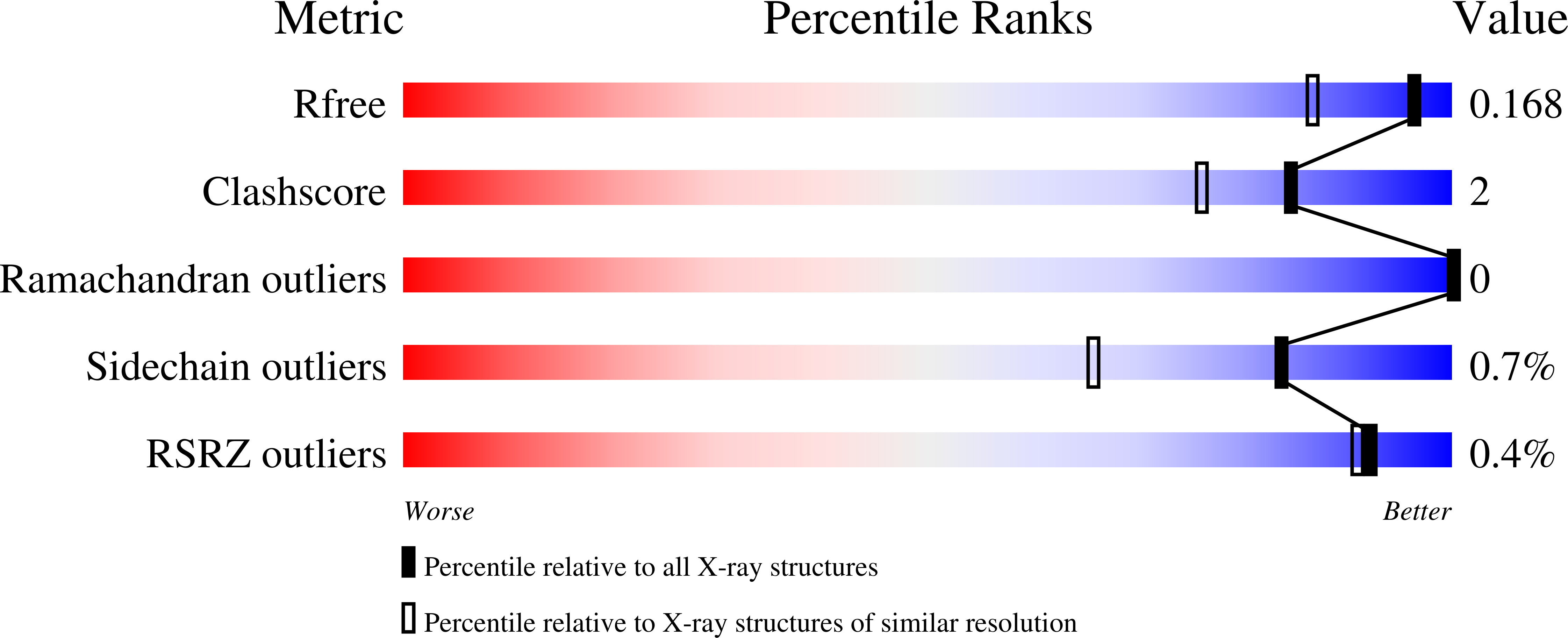
4NI3

Crystal Structure of GH29 family alpha-L-fucosidase from Fusarium graminearum in the closed form


Experimental Data Snapshot

  • Method: X-RAY DIFFRACTION
  • Resolution: 1.40 Å
  • R-Value Free: 0.167 
  • R-Value Work: 0.145 
  • R-Value Observed: 0.145 

wwPDB Validation   3D Report Full Report


Ligand Structure Quality Assessment 


This is version 2.1 of the entry. See complete history


Literature

Structure and Substrate Specificity of a Eukaryotic Fucosidase from Fusarium graminearum.

Cao, H.Walton, J.D.Brumm, P.Phillips, G.N.

(2014) J Biol Chem 289: 25624-25638

  • DOI: https://doi.org/10.1074/jbc.M114.583286
  • Primary Citation of Related Structures:  
    4NI3, 4PSP, 4PSR

  • PubMed Abstract: 

    The secreted glycoside hydrolase family 29 (GH29) α-L-fucosidase from plant pathogenic fungus Fusarium graminearum (FgFCO1) actively releases fucose from the xyloglucan fragment. We solved crystal structures of two active-site conformations, i.e. open and closed, of apoFgFCO1 and an open complex with product fucose at atomic resolution. The closed conformation supports catalysis by orienting the conserved general acid/base Glu-288 nearest the predicted glycosidic position, whereas the open conformation possibly represents an unreactive state with Glu-288 positioned away from the catalytic center. A flexible loop near the substrate binding site containing a non-conserved GGSFT sequence is ordered in the closed but not the open form. We also identified a novel C-terminal βγ-crystallin domain in FgFCO1 devoid of calcium binding motif whose homologous sequences are present in various glycoside hydrolase families. N-Glycosylated FgFCO1 adopts a monomeric state as verified by solution small angle x-ray scattering in contrast to reported multimeric fucosidases. Steady-state kinetics shows that FgFCO1 prefers α1,2 over α1,3/4 linkages and displays minimal activity with p-nitrophenyl fucoside with an acidic pH optimum of 4.6. Despite a retaining GH29 family fold, the overall specificity of FgFCO1 most closely resembles inverting GH95 α-fucosidase, which displays the highest specificity with two natural substrates harboring the Fucα1-2Gal glycosidic linkage, a xyloglucan-derived nonasaccharide, and 2'-fucosyllactose. Furthermore, FgFCO1 hydrolyzes H-disaccharide (lacking a +2 subsite sugar) at a rate 10(3)-fold slower than 2'-fucosyllactose. We demonstrated the structurally dynamic active site of FgFCO1 with flexible general acid/base Glu, a common feature shared by several bacterial GH29 fucosidases to various extents.


  • Organizational Affiliation

    From Rice University, Houston Texas 77005, Great Lakes Bioenergy Research Center, University of Wisconsin, Madison, Wisconsin 53706.


Macromolecules
Find similar proteins by:  (by identity cutoff)  |  3D Structure
Entity ID: 1
MoleculeChains Sequence LengthOrganismDetailsImage
Alpha-fucosidase GH29
A, B
585Fusarium graminearumMutation(s): 0 
Gene Names: FCO1
UniProt
Find proteins for J9UN47 (Gibberella zeae)
Explore J9UN47 
Go to UniProtKB:  J9UN47
Entity Groups  
Sequence Clusters30% Identity50% Identity70% Identity90% Identity95% Identity100% Identity
UniProt GroupJ9UN47
Sequence Annotations
Expand
  • Reference Sequence
Oligosaccharides

Help

Entity ID: 2
MoleculeChains Length2D Diagram Glycosylation3D Interactions
alpha-D-mannopyranose-(1-2)-alpha-D-mannopyranose-(1-2)-alpha-D-mannopyranose-(1-3)-beta-D-mannopyranose-(1-4)-2-acetamido-2-deoxy-beta-D-glucopyranose-(1-4)-2-acetamido-2-deoxy-beta-D-glucopyranose
C, D
6N-Glycosylation
Glycosylation Resources
GlyTouCan:  G01760ZU
GlyCosmos:  G01760ZU
GlyGen:  G01760ZU
Small Molecules
Ligands 4 Unique
IDChains Name / Formula / InChI Key2D Diagram3D Interactions
NAG
Query on NAG

Download Ideal Coordinates CCD File 
E [auth A],
F [auth A],
N [auth B]
2-acetamido-2-deoxy-beta-D-glucopyranose
C8 H15 N O6
OVRNDRQMDRJTHS-FMDGEEDCSA-N
TRS
Query on TRS

Download Ideal Coordinates CCD File 
I [auth A],
J [auth A],
Q [auth B],
R [auth B],
S [auth B]
2-AMINO-2-HYDROXYMETHYL-PROPANE-1,3-DIOL
C4 H12 N O3
LENZDBCJOHFCAS-UHFFFAOYSA-O
GOL
Query on GOL

Download Ideal Coordinates CCD File 
G [auth A]
H [auth A]
K [auth A]
M [auth A]
O [auth B]
G [auth A],
H [auth A],
K [auth A],
M [auth A],
O [auth B],
P [auth B],
T [auth B],
W [auth B]
GLYCEROL
C3 H8 O3
PEDCQBHIVMGVHV-UHFFFAOYSA-N
NA
Query on NA

Download Ideal Coordinates CCD File 
L [auth A],
U [auth B],
V [auth B]
SODIUM ION
Na
FKNQFGJONOIPTF-UHFFFAOYSA-N
Experimental Data & Validation

Experimental Data

  • Method: X-RAY DIFFRACTION
  • Resolution: 1.40 Å
  • R-Value Free: 0.167 
  • R-Value Work: 0.145 
  • R-Value Observed: 0.145 
  • Space Group: P 1
Unit Cell:
Length ( Å )Angle ( ˚ )
a = 53.511α = 105.61
b = 75.512β = 107.19
c = 80.367γ = 106.71
Software Package:
Software NamePurpose
PHENIXrefinement
PDB_EXTRACTdata extraction
MD2data collection
HKL-2000data reduction
HKL-2000data scaling
PHASERphasing

Structure Validation

View Full Validation Report


Ligand Structure Quality Assessment 


Entry History 

Deposition Data

Revision History  (Full details and data files)

  • Version 1.0: 2013-12-25
    Type: Initial release
  • Version 1.1: 2014-09-03
    Changes: Database references
  • Version 1.2: 2014-10-01
    Changes: Database references
  • Version 2.0: 2020-07-29
    Type: Remediation
    Reason: Carbohydrate remediation
    Changes: Atomic model, Data collection, Derived calculations, Structure summary
  • Version 2.1: 2023-09-20
    Changes: Data collection, Database references, Refinement description, Structure summary