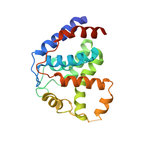
Crystal structure of PhnZ in complex with substrate reveals a di-iron oxygenase mechanism for catabolism of organophosphonates.
van Staalduinen, L.M., McSorley, F.R., Schiessl, K., Seguin, J., Wyatt, P.B., Hammerschmidt, F., Zechel, D.L., Jia, Z.(2014) Proc Natl Acad Sci U S A 111: 5171-5176
- PubMed: 24706911
- DOI: https://doi.org/10.1073/pnas.1320039111
- Primary Citation of Related Structures:
4MLM, 4MLN - PubMed Abstract:
The enzymes PhnY and PhnZ comprise an oxidative catabolic pathway that enables marine bacteria to use 2-aminoethylphosphonic acid as a source of inorganic phosphate. PhnZ is notable for catalyzing the oxidative cleavage of a carbon-phosphorus bond using Fe(II) and dioxygen, despite belonging to a large family of hydrolytic enzymes, the HD-phosphohydrolase superfamily. We have determined high-resolution structures of PhnZ bound to its substrate, (R)-2-amino-1-hydroxyethylphosphonate (2.1 Å), and a buffer additive, l-tartrate (1.7 Å). The structures reveal PhnZ to have an active site containing two Fe ions coordinated by four histidines and two aspartates that is strikingly similar to the carbon-carbon bond cleaving enzyme, myo-inositol-oxygenase. The exception is Y24, which forms a transient ligand interaction at the dioxygen binding site of Fe2. Site-directed mutagenesis and kinetic analysis with substrate analogs revealed the roles of key active site residues. A fifth histidine that is conserved in the PhnZ subclade, H62, specifically interacts with the substrate 1-hydroxyl. The structures also revealed that Y24 and E27 mediate a unique induced-fit mechanism whereby E27 specifically recognizes the 2-amino group of the bound substrate and toggles the release of Y24 from the active site, thereby creating space for molecular oxygen to bind to Fe2. Structural comparisons of PhnZ reveal an evolutionary connection between Fe(II)-dependent hydrolysis of phosphate esters and oxidative carbon-phosphorus or carbon-carbon bond cleavage, thus uniting the diverse chemistries that are found in the HD superfamily.
Organizational Affiliation:
Department of Biomedical and Molecular Sciences, Queen's University, Kingston, ON, Canada K7L 3N6.